. 2000 Mar;74(5):2084-93.
doi: 10.1128/jvi.74.5.2084-2093.2000.

Induction of the cellular E2F-1 promoter by the adenovirus E4-6/7 protein

Affiliations

Induction of the cellular E2F-1 promoter by the adenovirus E4-6/7 protein

J Schaley et al. J Virol. 2000 Mar.

Abstract

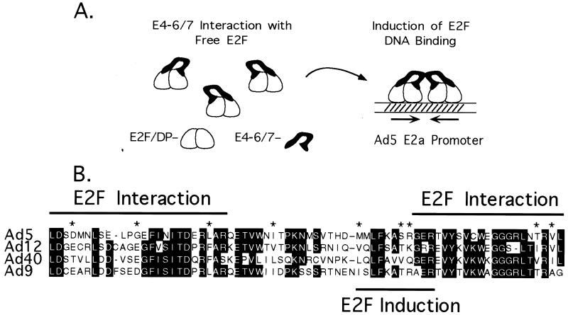
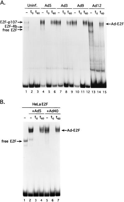
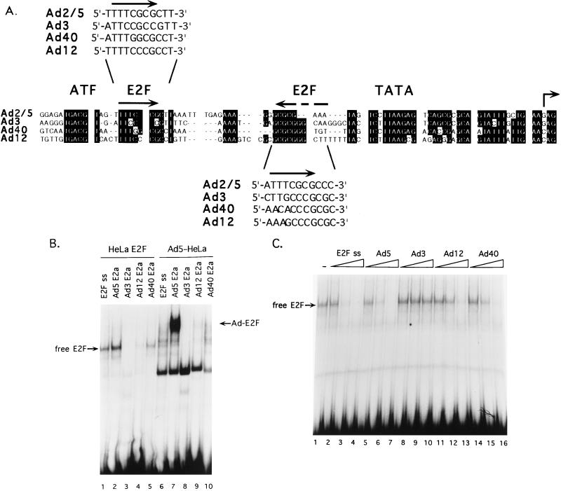
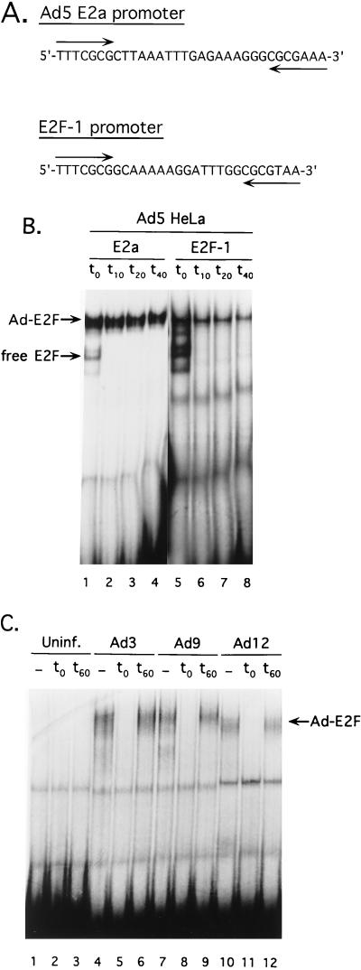
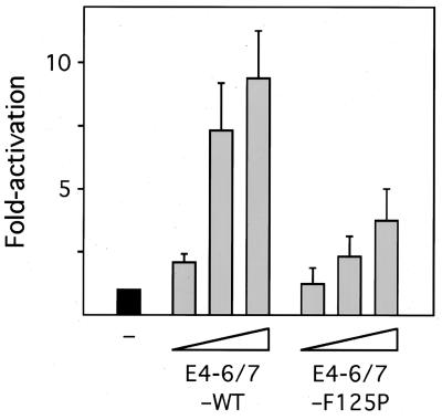
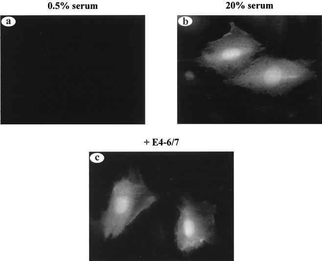
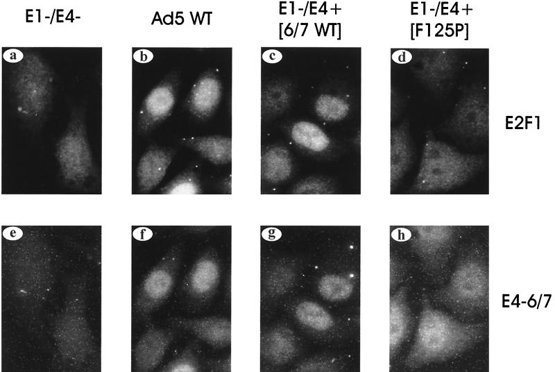
The adenovirus type 5 (Ad5) E4-6/7 protein interacts directly with different members of the E2F family and mediates the cooperative and stable binding of E2F to a unique pair of binding sites in the Ad5 E2a promoter region. This induction of E2F DNA binding activity strongly correlates with increased E2a transcription when analyzed using virus infection and transient expression assays. Here we show that while different adenovirus isolates express an E4-6/7 protein that is capable of induction of E2F dimerization and stable DNA binding to the Ad5 E2a promoter region, not all of these viruses carry the inverted E2F binding site targets in their E2a promoter regions. The Ad12 and Ad40 E2a promoter regions bind E2F via a single binding site. However, these promoters bind adenovirus-induced (dimerized) E2F very weakly. The Ad3 E2a promoter region binds E2F very poorly, even via a single binding site. A possible explanation of these results is that the Ad E4-6/7 protein evolved to induce cellular gene expression. Consistent with this notion, we show that infection with different adenovirus isolates induces the binding of E2F to an inverted configuration of binding sites present in the cellular E2F-1 promoter. Transient expression of the E4-6/7 protein alone in uninfected cells is sufficient to induce transactivation of the E2F-1 promoter linked to chloramphenicol acetyltransferase or green fluorescent protein reporter genes. Further, expression of the E4-6/7 protein in the context of adenovirus infection induces E2F-1 protein accumulation. Thus, the induction of E2F binding to the E2F-1 promoter by the E4-6/7 protein observed in vitro correlates with transactivation of E2F-1 promoter activity in vivo. These results suggest that adenovirus has evolved two distinct mechanisms to induce the expression of the E2F-1 gene. The E1A proteins displace repressors of E2F activity (the Rb family members) and thus relieve E2F-1 promoter repression; the E4-6/7 protein complements this function by stably recruiting active E2F to the E2F-1 promoter to transactivate expression.


Figures

FIG. 1
FIG. 1
(A) Model for Ad induction of E2F DNA binding. The left depicts interaction of the E4-6/7 protein with free E2F-DP heterodimers. The right depicts the induction of E2F DNA binding to the Ad5 E2a promoter region by E4-6/7 protein-mediated dimerization. The inverted E2F binding sites in the Ad5 E2a promoter are indicated by inverted arrows. (B) Alignment of E4-6/7 proteins from different Ad serotypes. E4 ORF7 of the Ad5 E4-6/7 protein is shown at the top in alignment with homologous sequences found in Ad12, Ad40, and Ad9. Dark letters indicate amino acid identities; conserved amino acid changes are indicated by asterisks across the top. The two regions in Ad5 E4-6/7 required for stable interaction with a E2F-DP heterodimer are indicated at the top of the sequence (termed E2F interaction), while the region of E4-6/7 that directs dimerization is shown at the bottom of the sequence (termed E2F induction).
FIG. 2
FIG. 2
Induction of E2F DNA binding to the Ad5 E2a promoter by different Ad serotypes. (A) Nuclear extracts from uninfected A549 cells (Uninf.; lanes 1 to 3) or A549 cells infected with Ad5 (lanes 4 to 6), Ad3 (lanes 7 to 9), Ad9 (lanes 10 to 12), or Ad12 (lanes 13 to 15) were used in gel mobility shift assays with a probe corresponding to the Ad5 E2a inverted E2F binding sites. The first lane of each set (lanes 1, 4, 7, 10, and 13) represents a binding reaction with no specific competitor DNA added. E2F complexes found in uninfected cells are indicated on the left, and the Ad-induced E2F complex is indicated on the right (Ad-E2F). In binding reactions shown in lanes 2, 5, 8, 11, and 14, a 500-fold molar excess of unlabeled DNA corresponding to an E2F single binding site (see Materials and Methods) was added to the binding reaction coincident with the addition of probe DNA. In binding reactions shown in lanes 3, 6, 9, 12, and 15, a 500-fold molar excess of unlabeled E2F single binding site DNA was added after a 60-min preincubation of the binding reaction with probe DNA. The binding reactions were incubated an additional 15 min before loading on the gel. (B) Bacterially expressed E4 ORF7 proteins were used in binding reactions with HeLa cell-free E2F activity. Lane 1 shows a binding reaction with HeLa cell cytoplasmic extract plus the Ad5 E2a probe. Free E2F binding activity is indicated on the left. In lanes 2 to 4, an Ad5 GST-E4-ORF7 fusion protein was added to the HeLa cell extract prior to the addition of probe DNA. In lanes 5 to 7, an Ad40 GST-E4-ORF7 fusion protein was added. Lanes 2 and 5 represent binding reactions without the addition of specific competitor DNA. In lanes 3 and 6, a 500-fold molar excess of unlabeled E2F binding site was added coincident with the probe as described for panel A. In lanes 4 and 7, a 500-fold molar excess of unlabeled E2F binding site was added after a 60-min binding reaction as described in for panel A.
FIG. 3
FIG. 3
E2a promoter regions of different Ad serotypes do not contain inverted E2F binding sites. (A) Alignment of the E2a promoter regions from different Ad serotypes. The ATF binding site, TATA box, and potential E2F sites are indicated at the top. Alignment of E2a promoters shows strong conservation of the ATF site, the TATA box, and start site region. The ATF-proximal and TATA-proximal potential E2F sites are shown in a 5′-to-3′ orientation in the insets above and below the sequence. (B) Binding reactions contained HeLa cell E2F and Ad5-induced E2F with probes corresponding to the E2a regions of different Ad serotypes. Lanes 1 to 5 show binding reactions with HeLa cell-free E2F activity and DNA probes corresponding to an E2F single-site (E2F ss) and E2a regions of Ad5, Ad3, Ad12 and Ad40, as indicated above the lanes. Lanes 6 to 10 show binding reactions with Ad5-infected HeLa cell nuclear extract with the same set of probes as described for lanes 1 to 5. The Ad-induced E2F complex is indicated in the right (Ad-E2F). The faster-migrating complex is not specifically competed by an E2F binding site and is an unknown binding activity that serves as an internal control for the integrity of each probe DNA. (C) The binding of HeLa cell free E2F to the E2a regions of different Ad serotypes was analyzed using competition binding assays. Lane 1 shows a binding reaction using HeLa cell-free E2F activity with an E2F single-site probe. Free E2F binding activity is indicated on the left. Lanes 2 to 16 show binding reactions where increasing molar concentrations (5-, 25-, and 125-fold molar excess to the probe DNA) of specific competitor DNAs were added coincident with the probe DNA. The cold competitors corresponded to an E2F single site identical to the probe DNA (lanes 2 to 4) and to E2a DNA of Ad5 (lanes 5 to 7), Ad3 (lanes 8 to 10), Ad12 (lanes 11 to 13), and Ad40 (lanes 14 to 16).
FIG. 4
FIG. 4
Ad induction of E2F binding to the cellular E2F-1 promoter. (A) Nucleotide sequence comparison of the Ad5 E2a promoter E2F binding sites and similar sites found in the cellular E2F-1 promoter. Inverted E2F binding sites are indicated by arrows below the sequences. (B) Binding of Ad-induced E2F to the E2a and E2F-1 binding sites. Lanes 1 and 5 show binding reactions using Ad5-infected HeLa cell extract with probes corresponding to Ad5 E2a (lane 1) or the cellular E2F-1 promoter E2F binding sites (lane 5). The positions of free E2F and the Ad-induced E2F (Ad-E2F) complexes are indicated on the left. Following a 60-min binding reaction, a 500-fold molar excess of unlabeled E2F single binding site was added, and aliquots of the binding reaction were removed and loaded on the gel at the times indicated above the lanes (10, 20, and 40 min after the addition of specific competitor DNA). (C) Different Ad serotypes induce E2F binding to the E2F-1 promoter. Nuclear extracts from uninfected (Uninf.) A549 cells or A549 cells infected with Ad3, Ad9, or Ad12 were incubated with the probe corresponding to the E2F-1 promoter. Lanes 1, 4, 7, and 10 show a binding reaction without the addition of specific competitor DNA. In lanes 2, 5, 8, and 11, a 500-fold molar excess of unlabeled E2F single binding site was added coincident with the probe. In lanes 3, 6, 9, and 12, a 500-fold molar excess of unlabeled E2F binding site was added after a 60-min binding reaction. The binding reactions were incubated an additional 15 min before loading on the gel. The position of the Ad-induced E2F complex (Ad-E2F) is indicated on the right.
FIG. 5
FIG. 5
Transactivation of the E2F-1 promoter by the E4-6/7 protein in HepG2 cells. HepG2 cells were transfected with a reporter vector containing the cellular E2F-1 promoter fused to CAT. The reporter vectors were transfected alone (−) or with increasing concentrations (2, 10, and 50 ng) of a vector expressing Ad2 E4-6/7-WT or E4-6/7-F125P. Following transfection, the cells were maintained in medium containing 0.5% serum for 24 h. The level of CAT activity in cellular extracts was then measured. The level of expression of the reporter vector alone is set at 1, and the results with E4-6/7 coexpression are indicated as fold activation relative to the basal level. The results represent the average of five experiments. Error bars indicate standard deviations.
FIG. 6
FIG. 6
Transactivation of the E2F-1 promoter by the E4-6/7 protein in REF52 cells. Quiescent REF52 cells were microinjected with the E2F-1–GFP reporter vector. Cells were coinjected with the E2F-1–GFP vector and an empty CMV vector (a and b) or coinjected with the E2F-1–GFP vector and an expression vector for the Ad2 E4-6/7 protein. In panels a and c, the cells were maintained in 0.5% serum; panel b, the cells were treated with medium containing 20% FBS. GFP activity was visualized 16 h after microinjection using a fluorescein filter on an Axiovert 135 microscope (Zeiss).
FIG. 7
FIG. 7
Induction of E2F-1 protein levels by the E4-6/7 protein in Ad-infected HeLa cells. HeLa cells were infected with wild-type Adenovirus (expressing E1A and E4-6/7; b and f) or a recombinant that lacks the E1 region and constitutively expresses E4-6/7-WT (c and g), E4-6/7-F125P (d and h), or an E1-lacking virus that also contains a mutation that disrupts the E4-6/7 protein (a and e). Cells were harvested at 8 h after infection, and E2F-1 (a to d) and E4-6/7 (e to h) were visualized by indirect immunofluorescence using antibodies specific to these products.

Similar articles

Cited by

References

    1. Adnane J, Shao Z, Robbins P D. The retinoblastoma susceptibility gene product represses transcription when directly bound to the promoter. J Biol Chem. 1995;270:8837–8843. - PubMed
    1. Blake M C, Azizkhan J C. Transcription factor E2F is required for efficient expression of the hamster dihydrofolate reductase gene in vitro and in vivo. Mol Cell Biol. 1989;9:4994–5002. - PMC - PubMed
    1. Botz J, Zerfass-Thome K, Spitkovsky D, Delius H, Vogt B, Eilers M, Hatzigeorgiou A, Jansen-Durr P. Cell cycle regulation of the murine cyclin E gene depends on an E2F binding site in the promoter. Mol Cell Biol. 1996;16:3401–3409. - PMC - PubMed
    1. Brehm A, Miska E A, McCance D J, Reid J L, Bannister A J, Kouzarides T. Retinoblastoma protein recruits histone deacetylase to repress transcription. Nature. 1998;391:597–601. - PubMed
    1. Bremner R, Cohen B L, Sopta M, Hamel P A, Ingles C J, Gallie B L, Phillips R A. Direct transcriptional repression by pRB and its reversal by specific cyclins. Mol Cell Biol. 1995;15:3256–3265. - PMC - PubMed

Publication types

MeSH terms

LinkOut - more resources