. 2000 Mar;74(5):2107-20.
doi: 10.1128/jvi.74.5.2107-2120.2000.

Roscovitine, a specific inhibitor of cellular cyclin-dependent kinases, inhibits herpes simplex virus DNA synthesis in the presence of viral early proteins

Affiliations

Roscovitine, a specific inhibitor of cellular cyclin-dependent kinases, inhibits herpes simplex virus DNA synthesis in the presence of viral early proteins

L M Schang et al. J Virol. 2000 Mar.

Abstract

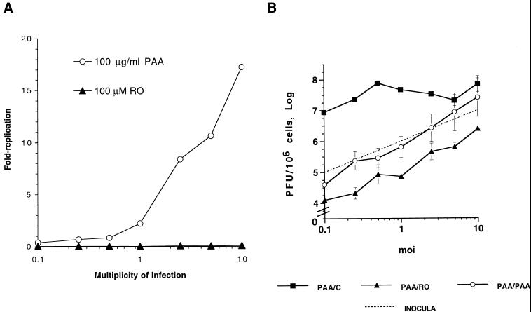
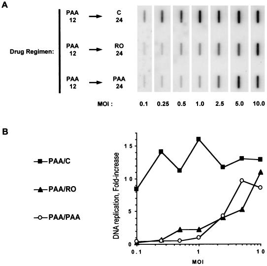
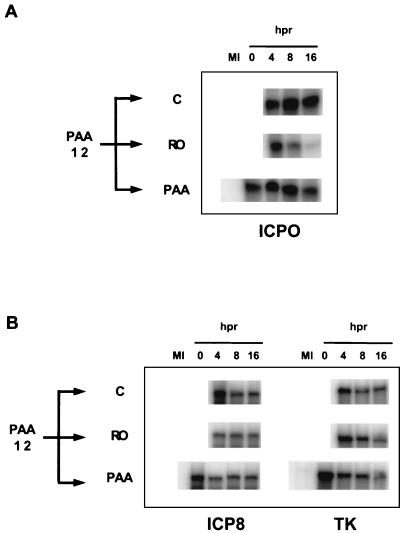
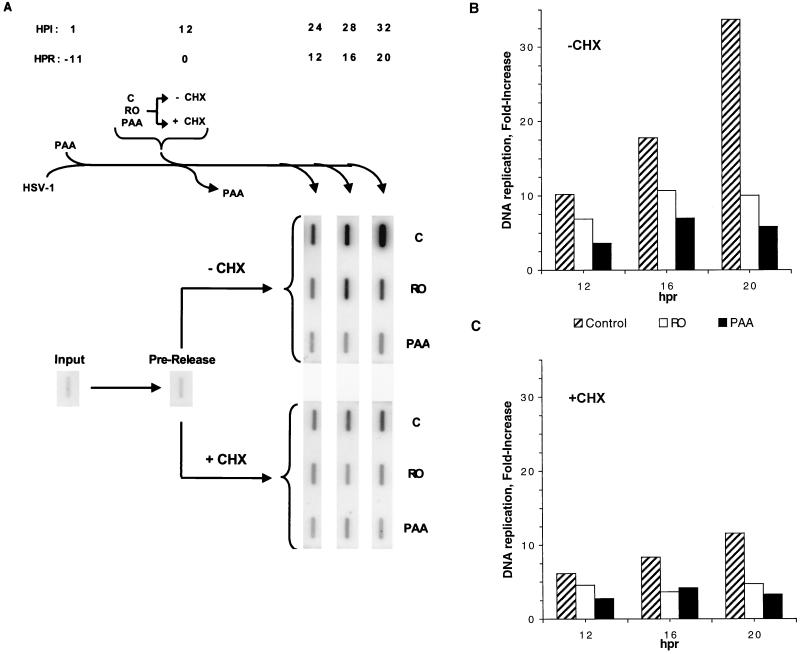
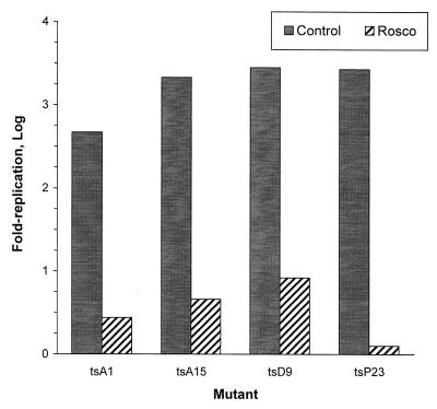
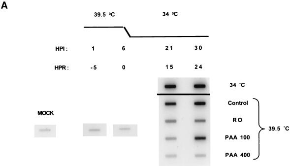
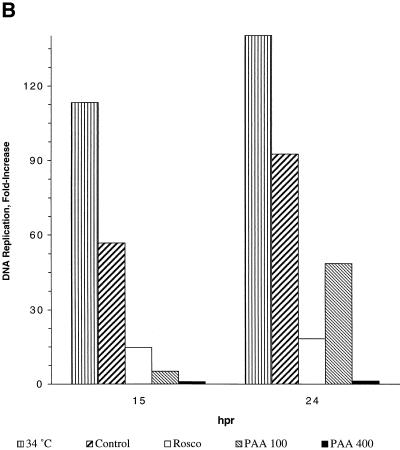
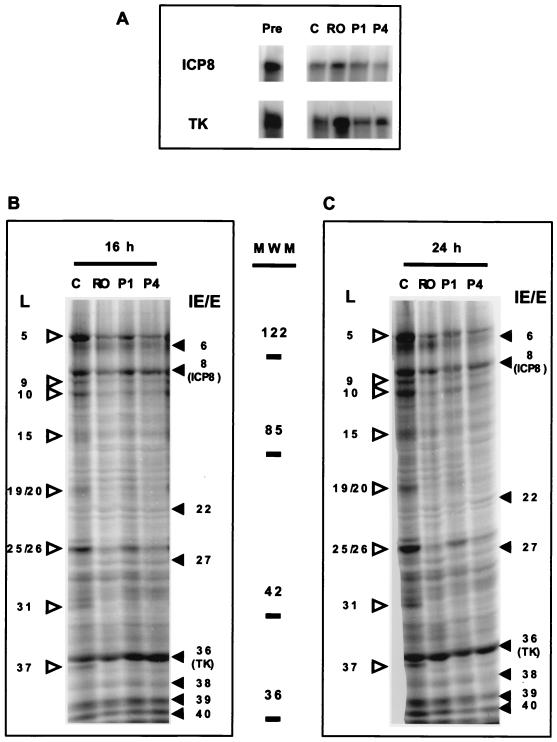
We have previously shown that two inhibitors specific for cellular cyclin-dependent kinases (cdks), Roscovitine (Rosco) and Olomoucine (Olo), block the replication of herpes simplex virus (HSV). Based on these results, we demonstrated that HSV replication requires cellular cdks that are sensitive to these drugs (L. M. Schang, J. Phillips, and P. A. Schaffer. J. Virol. 72:5626-5637, 1998). We further established that at least two distinct steps in the viral replication cycle require cdks: transcription of immediate-early (IE) genes and transcription of early (E) genes (L. M. Schang, A. Rosenberg, and P. A. Schaffer, J. Virol. 73:2161-2172, 1999). Since Rosco inhibits HSV replication efficiently even when added to infected cells at 6 h postinfection, we postulated that cdks may also be required for viral functions that occur after E gene expression. In the study presented herein, we tested this hypothesis directly by measuring the efficiency of viral replication, viral DNA synthesis, and expression of several viral genes during infections in which Rosco was added after E proteins had already been synthesized. Rosco inhibited HSV replication, and specifically viral DNA synthesis, when the drug was added at the time of release from a 12-h phosphonoacetic acid (PAA)-induced block in viral DNA synthesis. Inhibition of DNA synthesis was not a consequence of inhibition of expression of IE or E genes in that Rosco had no effect on steady-state levels of two E transcripts under the same conditions in which it inhibited viral DNA synthesis. Moreover, viral DNA synthesis was inhibited by Rosco even in the absence of protein synthesis. In a second series of experiments, the replication of four HSV mutants harboring temperature-sensitive mutations in genes essential for viral DNA replication was inhibited when Rosco was added at the time of shift-down from the nonpermissive to the permissive temperature. Viral DNA synthesis was inhibited by Rosco under these conditions, whereas expression of viral E genes was not affected. We conclude that cellular Rosco-sensitive cdks are required for replication of viral DNA in the presence of viral E proteins. This requirement may indicate that HSV DNA synthesis is functionally linked to transcription, which requires cdks, or that both viral transcription and DNA replication, independently, require viral or cellular factors activated by Rosco-sensitive cdks.


Figures

FIG. 1
FIG. 1
Effect of Rosco on HSV replication when added at 3-h intervals after infection. Vero cells were infected with 2.5 PFU of HSV-1 per cell. After 1 h of adsorption, the cells were washed and overlaid with medium containing no drug (control) or 100 μM Rosco. At 3, 6, 9, 12, 15, 18, and 21 h p.i., medium was removed and replaced with medium containing 100 μM Rosco (dotted lines, solid triangles). Changes from drug-free to Rosco-containing medium are indicated by the arrows. A second set of infected monolayers was left in drug-free medium (solid line, solid squares). At 1, 3, 6, 9, 12, 18, 21, and 24 h p.i., cultures were harvested and viral titers were determined by a standard plaque assay. Viral titers are plotted as a function of time p.i. Each time point indicates the average of two independent experiments.
FIG. 2
FIG. 2
The inhibition of HSV replication by Rosco added at the time of release from a 12-h PAA block is multiplicity dependent. (A) Vero cells were infected with HSV-1 at the indicated multiplicities of infection, washed, and overlaid with medium containing 100 μg of PAA per ml or 100 μM Rosco. Infected monolayers were harvested at 24 h p.i., and viral replication was calculated by dividing the viral titer at 24 h p.i. by the adsorbed PFU. (B) Vero cells were infected with HSV-1 at the indicated multiplicities, washed, and overlaid with medium containing 100 μg of PAA per ml. At 12 h p.i., medium was removed from infected monolayers and replaced with fresh medium containing no drug (PAA/C), 100 μg of PAA per ml (PAA/PAA), or 100 μM Rosco (PAA/RO). At 24 h after the change of medium, cells were harvested and viral titers were determined by a standard plaque assay. Viral yields at 24 h p.i. are plotted against the multiplicities of infection (moi). For reference, the dotted line indicates the number of PFU/106 cells in inocula at each multiplicity. The average and range of two independent experiments are shown for each time point.
FIG. 3
FIG. 3
The inhibition of HSV DNA synthesis by Rosco added at the time of release from a 12-h PAA block is multiplicity dependent. (A) Vero cells were infected at the indicated multiplicities (MOI) with HSV, washed, and overlaid with medium containing 100 μg of PAA per ml. At 12 h p.i. medium was removed and fresh medium containing no drug (PAA 12 → C 24), 100 μg of PAA per ml (PAA 12 → PAA 24), or 100 μM Rosco (PAA 12 → Ro 24) was added. At 24 h after the change of medium, cells were harvested and the amounts of viral DNA were determined by slot blot analysis. (B) After quantitation of the blots presented in panel A using a Molecular Dynamics PhosphorImager system, the fold increase in viral DNA replication was calculated by dividing the amount of viral DNA detected at 12 h postrelease by the amount detected immediately after infection and subtracting 1, such that a total block in viral DNA replication by the secondary drug would be indicated by 0-fold increase. The fold increase in DNA replication was then plotted against the multiplicity (MOI). The results from one of two repeat experiments are presented.
FIG. 4
FIG. 4
Effect of Rosco and PAA on the steady-state levels of selected viral transcripts when drug is added at the time of release from a 12-h PAA block. Vero cells were infected with 2 PFU of HSV per cell, and medium was replaced as described in the legend to Fig. 3B. Immediately before release (0) and at 4, 8, and 16 h after release (hpr) from the PAA block and addition of the secondary drug, cells were harvested and total RNA was extracted. RNA extracted from mock-infected cells served as a negative control (MI). Levels of ICP0 (IE), ICP8 (E), and TK (E) transcripts were determined by RNase protection assays.
FIG. 5
FIG. 5
Inhibition of HSV DNA synthesis by Rosco in the absence or presence of CHX added at the time of release from a 12-h PAA block. (A) Vero cells were infected with 2 PFU of HSV per cell, incubated in the presence of 100 μg of PAA per ml for 12 h, and released from the block as described in the legend to Fig. 3A, except that CHX alone or together with the secondary drug was added to one set of infected monolayers at the time of release (+ CHX). At 1 h p.i. (HPI) (−11 h postrelease [HPR]) immediately before release (0 h postrelease; 12 h p.i.), and at 12, 16, and 20 h postrelease (24, 28, and 32 h p.i., respectively), cells were harvested and total DNA was extracted. Levels of viral DNA were determined by slot blot hybridization at the indicated times. (B and C) Viral DNA in the blots in panel A were quantitated using a Molecular Dynamics PhosphorImager system, and the fold increase in viral DNA synthesis after release from the PAA block in the absence (B) (−CHX) or presence (C) (+CHX) of CHX was calculated by dividing the amounts of viral DNA at the indicated times postrelease by the amounts of viral DNA before release and subtracting 1, such that total inhibition of DNA replication by the secondary drug would be represented by 0-fold increase. The fold increase in DNA synthesis is plotted against hours postrelease (hpr). Data from one of two repeat experiments is presented.
FIG. 6
FIG. 6
Replication of four HSV ts mutants after the shift-down from the nonpermissive to the permissive temperature in the presence of Rosco. Vero cells were infected at the nonpermissive temperature with 2.5 PFU of the indicated HSV ts mutants per cell. At 6 h p.i., infected cultures were transferred to the permissive temperature in the absence of drug (Control) or in the presence of 100 μM Rosco. One culture infected with each ts mutant was harvested immediately before the shift-down. At 24 h after the shift-down, the remaining infected monolayers were harvested and viral replication was monitored by a standard plaque assay. Fold viral replication after release was determined by dividing the titers at 24 h postrelease by the titers measured before release and subtracting 1.
FIG. 7
FIG. 7
HSV DNA synthesis after the shift-down from the nonpermissive to the permissive temperature in the presence of Rosco added at the time of the shift-down. (A) Vero cells were infected with HSV tsA15 at the nonpermissive temperature and shifted down in the absence of drug or in the presence of 100 μM Rosco or 100 or 400 μg of PAA per ml (Control, RO, PAA100, and PAA400, respectively), as described in the text. At 1 h p.i. (HPI) (−5 h postrelease [HPR]), immediately before release (6 h pi/0 hpr) and at 15 and 24 h after release (21 or 30 h p.i., respectively), cells were harvested and DNA was extracted. DNA was also extracted from mock-infected cells for comparison (MOCK). Levels of viral DNA synthesis were determined by slot blot analysis. For comparison, a set of infected monolayers was incubated at the permissive temperature throughout the experiment. (B) The slot blots shown in panel A were quantitated using the ImageQuant software package (Molecular Dynamics), and the fold increase in DNA synthesis was calculated by dividing the amount of viral DNA at a given time point by the amount of viral DNA detected immediately before release and subtracting 1, such that complete inhibition of viral DNA replication after release would be indicated by 0-fold increase. The fold increase in DNA replication is plotted against hours postrelease (hpr). Data from one of two repeat experiments are presented.
FIG. 7
FIG. 7
HSV DNA synthesis after the shift-down from the nonpermissive to the permissive temperature in the presence of Rosco added at the time of the shift-down. (A) Vero cells were infected with HSV tsA15 at the nonpermissive temperature and shifted down in the absence of drug or in the presence of 100 μM Rosco or 100 or 400 μg of PAA per ml (Control, RO, PAA100, and PAA400, respectively), as described in the text. At 1 h p.i. (HPI) (−5 h postrelease [HPR]), immediately before release (6 h pi/0 hpr) and at 15 and 24 h after release (21 or 30 h p.i., respectively), cells were harvested and DNA was extracted. DNA was also extracted from mock-infected cells for comparison (MOCK). Levels of viral DNA synthesis were determined by slot blot analysis. For comparison, a set of infected monolayers was incubated at the permissive temperature throughout the experiment. (B) The slot blots shown in panel A were quantitated using the ImageQuant software package (Molecular Dynamics), and the fold increase in DNA synthesis was calculated by dividing the amount of viral DNA at a given time point by the amount of viral DNA detected immediately before release and subtracting 1, such that complete inhibition of viral DNA replication after release would be indicated by 0-fold increase. The fold increase in DNA replication is plotted against hours postrelease (hpr). Data from one of two repeat experiments are presented.
FIG. 8
FIG. 8
Expression of E gene products by tsA15 in the presence of Rosco added at the time of the shift-down. (A) Vero cells were infected with tsA15 at the nonpermissive temperature and shifted down to the permissive temperature as described in the legend to Fig. 7. Immediately before release (Pre) or 16 h after the shift-down in the absence of drug or in the presence of 100 μM Rosco or 100 or 400 μg of PAA per ml (C, RO, P1, and P4, respectively), cells were harvested and RNA was extracted. Steady-state levels of the transcripts of the genes encoding ICP8 or TK were evaluated by RNase protection assays. (B) Vero cells were infected with tsA15 at the nonpermissive temperature and shifted down to the permissive temperature as described for panel A, except that methionine-free medium supplemented with 50 μCi of [35S]methionine per ml and the indicated drugs (C, RO, P1, or P4) were added at the time of the shift-down. Infected cells were harvested at 16 h p.i., and viral proteins were resolved in a sodium dodecyl sulfate-polyacrylamide gel. Molecular weights (in thousands) are indicated on the right. The ICP nomenclature was used, such that VP16 is designated ICP25/26. HSV late proteins (γ1 and γ2) are indicated on the left of the gel by open arrowheads. Viral IE and E proteins are indicated on the right of the gel by solid arrowheads. (C) Infected cells were infected at the nonpermissive temperature, shifted down, and labeled with [5S]methionine as described in the legend to panel B, except that the cells were harvested at 24 h after the shift-down. Molecular weights, ICP nomenclature, and HSV IE, E, and L proteins (γ1 and γ2) are labeled as in panel B.

Similar articles

Cited by

References

    1. Abraham R T, Acquarone M, Andersen A, Asensi A, Belle R, Berger F, Bergounioux C, Brunn G, Buquet-Fagot C, Fagot D, et al. Cellular effects of olomoucine, an inhibitor of cyclin-dependent kinases. Biol Cell. 1995;83:105–120. - PubMed
    1. Alessi F, Quarta S, Savio M, Riva F, Rossi L, Stivala L, Scovassi A, Meijer L, Prosperi E. The cyclin-dependent kinase inhibitors olomoucine and roscovitine arrest human fibroblasts in G1 phase by specific inhibition of CDK2 kinase activity. Exp Cell Res. 1998;245:8–18. - PubMed
    1. Becker Y, Asher Y, Cohen Y, Weinberg-Zahlering E, Shlomai J. Phosphonoacetic acid-resistant mutants of herpes simplex virus: effect of phosphonoacetic acid on virus replication and in vitro deoxyribonucleic acid synthesis in isolated nuclei. Antimicrob Agents Chemother. 1997;11:919–922. - PMC - PubMed
    1. Beijersbergen R L, Carlee L, Kerkhoven R M, Bernards R. Regulation of the retinoblastoma protein-related p107 by G1 cyclin complexes. Genes Dev. 1995;9:1340–1353. - PubMed
    1. Bohemer P E, Lehman I. Herpes simplex virus DNA replication. Annu Rev Biochem. 1997;66:347–384. - PubMed

Publication types

MeSH terms

LinkOut - more resources