. 2000 Mar;74(5):2203-9.
doi: 10.1128/jvi.74.5.2203-2209.2000.

Cellular receptor traffic is essential for productive duck hepatitis B virus infection

Affiliations

Cellular receptor traffic is essential for productive duck hepatitis B virus infection

K M Breiner et al. J Virol. 2000 Mar.

Abstract

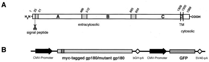
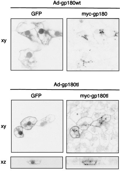
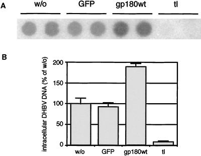
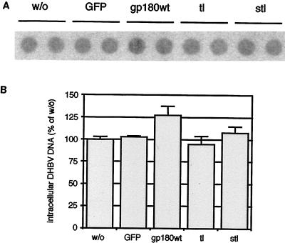
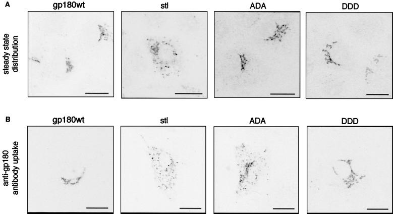
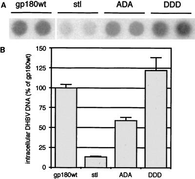
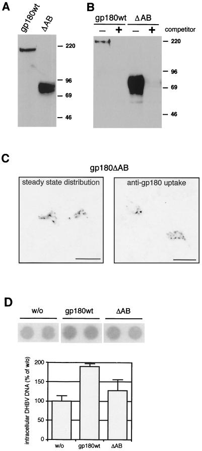
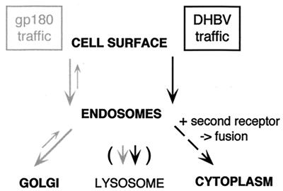
We have investigated the mechanism of duck hepatitis B virus (DHBV) entry into susceptible primary duck hepatocytes (PDHs), using mutants of carboxypeptidase D (gp180), a transmembrane protein shown to act as the primary cellular receptor for avian hepatitis B virus uptake. The variant proteins were abundantly produced from recombinant adenoviruses and tested for the potential to functionally outcompete the endogenous wild-type receptor. Overexpression of wild-type gp180 significantly enhanced the efficiency of DHBV infection in PDHs but did not affect ongoing DHBV replication, an observation further supporting gp180 receptor function. A gp180 mutant deficient for endocytosis abolished DHBV infection, indicating endocytosis to be the route of hepadnaviral entry. With further gp180 variants, carrying mutations in the cytoplasmic domain and characterized by an accelerated turnover, the ability of gp180 to function as a DHBV receptor was found to depend on a wild-type-like sorting phenotype which largely avoids transport toward the endolysosomal compartment. Based on these data, we propose a model in which a distinct intracellular DHBV traffic to the endosome, but not beyond, is a prerequisite for completion of viral entry, i.e., for fusion and capsid release. Furthermore, the deletion of the two enzymatically active carboxypeptidase domains of gp180 did not lead to a loss of receptor function.


Figures

FIG. 1
FIG. 1
(A) Schematic representation of duck carboxypeptidase D (gp180). Three homologous extracytosolic domains (A to C) are preceded by a signal peptide and followed by a transmembrane (TM) region and a short cytosolic domain. Numbers at the top indicate amino acid positions (13, 18). (B) Schematic representation of the expression cassettes, inserted in reverse direction into the deleted E1 region of the recombinant adenovirus vector (for details, see Materials and Methods). bGH-pA and SV40-pA, bovine growth hormone and simian virus 40 poly(A) sites.
FIG. 2
FIG. 2
Selective visualization in duck hepatocytes of Myc-tagged mutant gp180 expressed from recombinant adenoviruses. Primary hepatocytes were infected with recombinant adenoviruses encoding GFP plus Myc-tagged gp180 (Ad-gp180wt) or GFP plus a Myc-tagged gp180 mutant lacking the gp180 cytoplasmic domain (Ad-gp180tl). On day 4 postinfection, cells were fixed and stained with an anti-Myc antibody (tetramethylrhodamine), and GFP- and anti-Myc-related fluorescence was analyzed by confocal laser scanning microscopy (xy, in the plane of the culture; xz, perpendicular to the culture).
FIG. 3
FIG. 3
Expression of gp180 lacking the cytoplasmic domain blocks DHBV infection. PDHs were transduced with recombinant adenoviruses encoding GFP (GFP), GFP plus gp180 (gp180wt), or GFP plus gp180tl (tl) and after further 4 days infected with DHBV. w/o, without adenovirus transduction. Four days later, the cells were harvested and assayed by DNA dot blotting for intracellular DHBV DNA (A, duplicate determinations). (B) Relative quantification of radioactive signals. Error bars indicate the deviation between duplicate experiments.
FIG. 4
FIG. 4
Expression of mutant gp180 has no significant influence on ongoing DHBV replication. PDHs, prepared from a DHBV-positive duckling, were infected with recombinant adenoviruses encoding GFP only (GFP) or GFP plus gp180 (gp180wt) or deletion mutants tl and stl. w/o, control without adenovirus infection. At day 6 after adenovirus transduction, the cells were harvested and assayed by DNA dot blot for intracellular DHBV DNA (A, two parallel experiments). (B) Relative quantification of radioactive signals. Error bars indicate the deviation between duplicate measurements.
FIG. 5
FIG. 5
Changes in localization and endocytotic sorting of gp180 mutants carrying mutations in the cytoplasmic domain. HuH7 cells were transfected with plasmids encoding gp180wt or the indicated mutants of gp180 (Table 1). One day later, cells were immunostained with anti-gp180 antiserum and analyzed for gp180 localization by confocal laser scanning microscopy (A). Another set of cells was incubated with anti-gp180 antibodies for 2 h prior to fixation, staining with a fluorescent secondary antibody, and confocal microscopy (B). For a summary of mutant gp180 distribution, see Table 1. The scale bars represent 20 μm.
FIG. 6
FIG. 6
Disruption of intracellular sorting of gp180 influences DHBV infection. PDHs, preinfected with recombinant adenoviruses encoding gp180wt or the indicated mutants of gp180 were superinfected with DHBV. After further 4 days, the cells were harvested and assayed by DNA dot blotting for intracellular DHBV DNA (A, duplicate measurements). (B) Quantification of relative radioactive signals. Error bars indicate the deviation between two parallel experiments.
FIG. 7
FIG. 7
The enzymatically active domains A and B of gp180 are not essential for DHBV binding and receptor function. (A) Anti-gp180 immunoblot of lysates from HuH7 cells, transfected with plasmids encoding gp180wt or gp180ΔAB (ΔAB; see Fig. 1A and Materials and Methods). Numbers on the right indicate molecular masses of marker proteins (in kilodaltons). (B) Anti-gp180 immunoblot detecting gp180 or gp180ΔAB bound to a pre-S matrix in the absence (−) or presence (+) of free pre-S polypeptide as a competitor (2). (C) Localization and sorting of gp180ΔAB in transfected HuH7 cells. Cells were treated and analyzed by confocal microscopy as described for Fig. 5. The scale bars represent 20 μm. (D) Influence on DHBV infection. PDHs were infected with DHBV after transduction with recombinant adenoviruses encoding gp180wt or gp180ΔAB (ΔAB). w/o, without adenovirus transduction. Cells were assayed by DNA dot blotting for intracellular DHBV DNA as described for Fig. 3 and 6.
FIG. 8
FIG. 8
Model of cellular gp180 traffic and DHBV entry. DHBV-complexed gp180 is arrested in the endosome to allow interaction with the second receptor and membrane fusion.

References

    1. Breiner K M. Carboxypeptidase D (gp180): Rezeptor, Transrezeptor und Signalmolekül für Vogelhepatitis B Viren. Ph.D. thesis. Heidelberg, Germany: University of Heidelberg; 1998.
    1. Breiner K M, Urban S, Schaller H. Carboxypeptidase D (gp180), a Golgi-resident protein, functions in the attachment and entry of avian hepatitis B viruses. J Virol. 1998;72:8098–8104. - PMC - PubMed
    1. DeMeyer S, Gong J Z, Suwandhi W, van Pelt J, Soumillon A, Yap S H. Organ and species specificity of hepatitis B virus (HBV) infection: a review of literature with a special reference to preferential attachment of HBV to human hepatocytes. J Viral Hepat. 1997;4:145–153. - PubMed
    1. Dittie A S, Thomas L, Thomas G, Tooze S A. Interaction of furin in immature secretory granules from neuroendocrine cells with the AP-1 adaptor complex is modulated by casein kinase II phosphorylation. EMBO J. 1997;16:4859–4870. - PMC - PubMed
    1. Eng F J, Novikova E G, Kuroki K, Ganem D, Fricker L D. gp180, a protein that binds duck hepatitis B virus particles, has metallocarboxypeptidase D-like enzymatic activity. J Biol Chem. 1998;273:8382–8388. - PubMed

Publication types

Substances