. 2000 Mar;74(5):2323-32.
doi: 10.1128/jvi.74.5.2323-2332.2000.

Rotavirus infection induces an increase in intracellular calcium concentration in human intestinal epithelial cells: role in microvillar actin alteration

Affiliations

Rotavirus infection induces an increase in intracellular calcium concentration in human intestinal epithelial cells: role in microvillar actin alteration

J P Brunet et al. J Virol. 2000 Mar.

Abstract

Rotaviruses, which infect mature enterocytes of the small intestine, are recognized as the most important cause of viral gastroenteritis in young children. We have previously reported that rotavirus infection induces microvillar F-actin disassembly in human intestinal epithelial Caco-2 cells (N. Jourdan, J. P. Brunet, C. Sapin, A. Blais, J. Cotte-Laffitte, F. Forestier, A. M. Quero, G. Trugnan, and A. L. Servin, J. Virol. 72:7228-7236, 1998). In this study, to determine the mechanism responsible for rotavirus-induced F-actin alteration, we investigated the effect of infection on intracellular calcium concentration ([Ca(2+)](i)) in Caco-2 cells, since Ca(2+) is known to be a determinant factor for actin cytoskeleton regulation. As measured by quin2 fluorescence, viral replication induced a progressive increase in [Ca(2+)](i) from 7 h postinfection, which was shown to be necessary and sufficient for microvillar F-actin disassembly. During the first hours of infection, the increase in [Ca(2+)](i) was related only to an increase in Ca(2+) permeability of plasmalemma. At a late stage of infection, [Ca(2+)](i) elevation was due to both extracellular Ca(2+) influx and Ca(2+) release from the intracellular organelles, mainly the endoplasmic reticulum (ER). We noted that at this time the [Ca(2+)](i) increase was partially related to a phospholipase C (PLC)-dependent mechanism, which probably explains the Ca(2+) release from the ER. We also demonstrated for the first time that viral proteins or peptides, released into culture supernatants of rotavirus-infected Caco-2 cells, induced a transient increase in [Ca(2+)](i) of uninfected Caco-2 cells, by a PLC-dependent efflux of Ca(2+) from the ER and by extracellular Ca(2+) influx. These supernatants induced a Ca(2+)-dependent microvillar F-actin alteration in uninfected Caco-2 cells, thus participating in rotavirus pathogenesis.


Figures
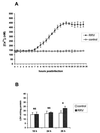
FIG. 1
FIG. 1
(A) [Ca2+]i in RRV-infected Caco-2 cells. Monolayers of 14- to 16-day-old Caco-2 cells were infected with RRV at an MOI of 10 PFU per cell and trypsinized at indicated times p.i. to measure [Ca2+]i by quin2 fluorescence. Each point corresponds to the mean ± standard deviation of six independent measurements. (B) Time course of cell membrane integrity. Infected and mock-infected cells were assayed at indicated times p.i. for LDH release. Values are means ± standard deviations from 10 experiments. Statistical differences between control and infected cells were determined by Student's t test. NS, not significantly different; ∗, P < 0.01.
FIG. 2
FIG. 2
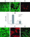
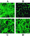
Microvillar F-actin disassembly induced by RRV infection (A to C). At 18 h p.i., RRV-infected (B) and mock-infected (A) Caco-2 cells were fixed, permeabilized, and stained with fluorescein-phalloidin, which binds to F-actin. RRV proteins were immunostained with polyclonal anti-group A rotavirus antibody and rhodamine-labeled anti-rabbit immunoglobulin G antibody. A horizontal section was generated by CLSM at the apex of the cells. (C) RRV staining in the same cells as in panel B. Treatment of infected cells with EGTA and BAPTA-AM totally abolishes the RRV-induced [Ca2+]i rise (D). Infected Caco-2 cells were treated at 15 h p.i. for 3 h with 3 mM EGTA and 75 μM BAPTA-AM. At 18 h p.i., cells were trypsinized to measure [Ca2+]i by quin2 fluorescence. Values are means ± standard deviations from three experiments. Statistical differences between control and infected cells were determined by Student's t test. NS, not significantly different; ∗, P < 0.01. EGTA and BAPTA-AM treatment totally protects Caco-2 cells from RRV-induced microvillar F-actin alteration (E and F). (E) Microvillar F-actin staining in treated 18 h p.i.-infected cells; (F) RRV staining in the same cells as in panel E; (G) microvillar F-actin disorganization in Caco-2 cells stained with fluorescein-phalloidin after treatment for 7 min with 10 μM ionomycin. Bars, 10 μm.
FIG. 3
FIG. 3
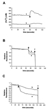
Determination of plasma membrane permeability to Ca2+ in RRV-infected Caco-2 cells. At indicated times p.i., Caco-2 cells were trypsinized, and cell suspensions were loaded with quin2-AM for measurements of [Ca2+]i. (A) Evaluation of Ca2+ permeability of plasmalemma by the change in [Ca2+]i induced by the addition of 5 mM CaCl2 to the EM (arrows), which initially contained 1.8 mM Ca2+, in 18 h p.i.-infected cells (a) and mock-infected cells (b). (B and C) Determination of plasmalemma permeability to Ca2+ by using Mn2+ as a substitute for Ca2+. The quenching of quin2 fluorescence induced by addition of 0.5 mM Mn2+ (arrows) was measured at an excitation wavelength of 366 nm. Relative fluorescence corresponds to the normalized fluorescence, taking initial values as maximal and Triton X-100 (T) values as minimal. (B) Results for 6 h p.i.-infected (a) and mock-infected (b) cells; (C) 18 h p.i.-infected (a) and mock-infected (b) cells. Representative traces of series of 10 experiments are shown.
FIG. 4
FIG. 4
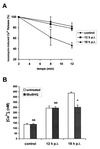
Ca2+ release from the internal stores at a late stage of RRV infection of Caco-2 cells. (A) Ca2+ efflux from ionomycin-sensitive stores in RRV-infected Caco-2 cells at indicated times p.i. and in mock-infected cells. The peak change in [Ca2+]i occurred following addition of ionomycin after 2, 8, and 12 min of suspension of quin2-AM-loaded cells in Ca2+-free EM containing 100 μM EGTA. Each point represents mean ± standard deviation of the net change in [Ca2+]i relative to the 2-min values from three independent experiments. (B) Effect of tBuBHQ on [Ca2+]i increase induced by RRV infection of Caco-2 cells. tBuBHQ (50 μM) was added to Caco-2 cells 1 h before trypsinization and quin2-AM loading. [Ca2+]i was measured in treated or untreated mock-infected and RRV-infected cells at indicated times p.i. Values are means ± standard deviations from six experiments. Statistical differences between untreated and tBuBHQ treated cells were determined by Student's t test. NS, not significantly different; ∗, P < 0.01.
FIG. 5
FIG. 5
At a late stage of RRV infection, [Ca2+]i increase in Caco-2 cells partially depends on a PLC mechanism. U-73122, a PLC inhibitor, or U-73343, a close analog, was added to Caco-2 cells at time zero. Mock-infected and RRV-infected cells were trypsinized at indicated times p.i. to measure [Ca2+]i by quin2 fluorescence. Values are means ± standard deviations from at least six experiments. Statistical differences between control and U-73122- or U-73343-treated cells were determined by Student's t test. NS, not significantly different; ∗, P < 0.01.
FIG. 6
FIG. 6
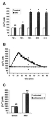
Effect of culture supernatants of RRV-infected Caco-2 on [Ca2+]i in uninfected Caco-2 cells. Caco-2 cells were infected with RRV at an MOI of 10 PFU/cell. (A) Peak values in [Ca2+]i were measured after the addition to 2 × 10⁶ quin2-AM loaded uninfected cells of supernatants of control or RRV-infected cells, collected at indicated times p.i. Values are means ± standard deviations from at least three to six experiments (see text). Statistical differences between peak values in [Ca2+]i of infected and control cells were determined by Student's t test. NS, not significantly different; ∗, P < 0.01. (B) Effect of heated or nonheated 18 h p.i.-infected supernatants on [Ca2+]i in uninfected Caco-2 cells. Supernatants of control cells (a), 18 h p.i.-infected cells (b), or 18 h p.i.-infected cells incubated at 95°C for 5 min (c) were added to uninfected Caco-2 cells immediately before [Ca2+]i measurements. Representative traces from three to six independent experiments are shown. (C) Effect of actinomycin D on the increase in [Ca2+]i induced by 18 h p.i.-infected cell supernatants. Supernatants issued from mock-infected or RRV-infected cells not treated or treated with actinomycin D (10 μg/ml) were added to quin2-loaded Caco-2 cells for [Ca2+]i measurements. Values are means ± standard deviations from three experiments. Statistical differences between [Ca2+]i of Caco-2 cells in the presence of treated and untreated supernatants were determined by Student's t test. NS, not significantly different.
FIG. 7
FIG. 7
Origin of the Ca2+ implicated in the [Ca2+]i increase induced by supernatants of RRV-infected Caco-2 cells. (A) Quin2-AM loaded Caco-2 cells, not treated (b) or treated with 3 mM EGTA a few seconds before the addition of 18 h p.i.-infected supernatants (a); (B) Caco-2 cells, not treated (b) or treated with 50 μM tBuBHQ 10 min before the addition of 18 h p.i.-infected supernatants (a); (C) Caco-2 cells, not treated (b) or treated with 3 mM EGTA and 50 μM tBuBHQ before the addition of 18 h p.i.-infected supernatants (a); (D) Caco-2 cells, treated with 50 μM U-73122 (a) or 50 μM U-73343 (b) 10 min before the addition of 18 h p.i.-infected supernatants. Representative traces from three independent experiments are shown.
FIG. 8
FIG. 8
Effect of supernatants of RRV-infected Caco-2 cells on microvillar F-actin organization of uninfected Caco-2 cells. At indicated times after the addition of 18 h p.i.-infected or mock-infected cell supernatants, Caco-2 cells were fixed, permeabilized, and stained with fluorescein-phalloidin. A horizontal section was generated by CLSM at the apex of the cells. F-actin staining 40 min after the addition of supernatants of mock-infected cells (A) or infected cells (B). (C) Normal F-actin pattern 90 min after the addition of supernatants of infected cells. Treatment of Caco-2 cells with 3 mM EGTA and 50 μM tBuBHQ (Fig. 7) prevented microvillar F-actin disorganization induced by supernatants of 18 h p.i.-infected cells (D). Bar, 10 μm.
FIG. 9
FIG. 9
A model depicting the mechanisms of [Ca2+]i increase in a monolayer of RRV-infected Caco-2 cells as a function of the stage of infection. At an early stage of infection (from 7 to 12 h p.i.), [Ca2+]i elevation is due only to extracellular Ca2+ entry and is partially compensated for by the activation of regulatory systems such as the Ca2+-ATPase pump of the ER. At a late stage of infection, the [Ca2+]i rise is more important in RRV-infected cells and induces microvillar F-actin disassembly from 18 h p.i. As of 15 h p.i., viral proteins or peptides released in extracellular medium from RRV-infected cells activate PLC in uninfected Caco-2 cells, through the interaction with a surface receptor. [Ca2+]i increases by an IP3-dependent mobilization of Ca2+ from the ER and by plasmalemma Ca2+ influx. This [Ca2+]i rise also leads to microvillar F-actin disassembly. PIP2, phosphatidylinositol 4,5-biphosphate; V, viroplasm.

Similar articles

Cited by

References

    1. Achler C, Filmer D, Merte C, Drenckhahn D. Role of microtubules in polarized delivery of apical membrane proteins to the brush border of the intestinal epithelium. J Cell Biol. 1989;109:179–189. - PMC - PubMed
    1. Ball J M, Tian P, Zeng C Q-Y, Morris A P, Estes M K. Age-dependent diarrhea induced by a rotaviral nonstructural glycoprotein. Science. 1996;272:101–104. - PubMed
    1. Berridge M J. Inositol trisphosphate and calcium signalling. Nature. 1993;361:315–325. - PubMed
    1. Bishop R F, Davidson G P, Holmes I H, Ruck B J. Virus particles in epithelial cells of duodenal mucosa from children with acute non-bacterial gastroenteritis. Lancet. 1973;ii:1281–1283. - PubMed
    1. Bleasdale J E, Thakur N R, Gremban R S, Bundy G L, Fitzpatrick F A, Smith R J, Bunting S. Selective inhibition of receptor-coupled phospholipase C-dependent processes in human platelets and polymorphonuclear neutrophils. J Pharmacol Exp Ther. 1990;255:756–768. - PubMed

Publication types