. 2000 Mar;74(5):2372-82.
doi: 10.1128/jvi.74.5.2372-2382.2000.

A chimeric protein containing the N terminus of the adeno-associated virus Rep protein recognizes its target site in an in vivo assay

Affiliations

A chimeric protein containing the N terminus of the adeno-associated virus Rep protein recognizes its target site in an in vivo assay

T Cathomen et al. J Virol. 2000 Mar.

Abstract

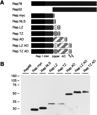
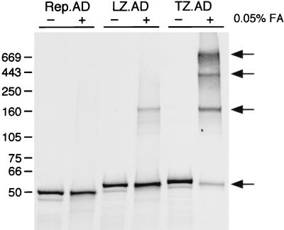
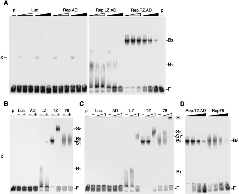
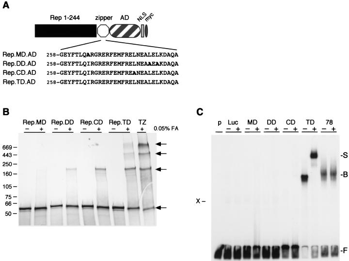
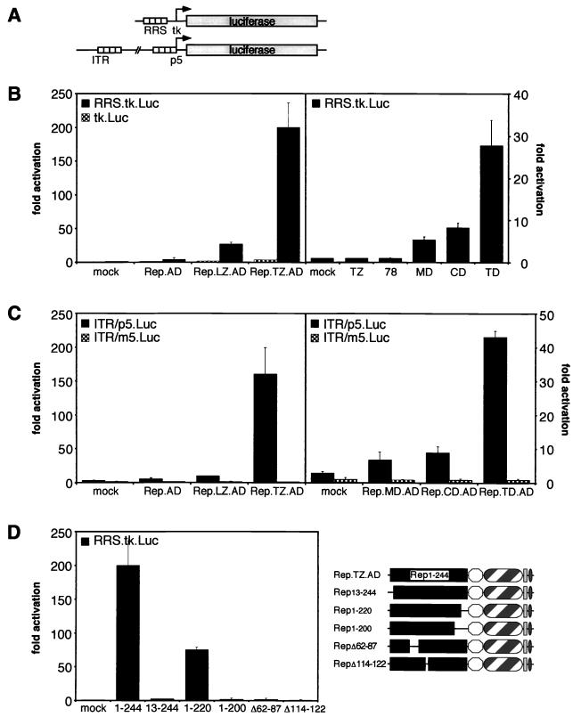
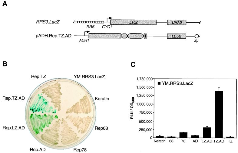
The Rep78 and Rep68 proteins of adeno-associated virus (AAV) type 2 are involved in DNA replication, regulation of gene expression, and targeting site-specific integration. They bind to a specific Rep recognition sequence (RRS) found in both the viral inverted terminal repeats and the AAVS1 integration locus on human chromosome 19. Previous in vitro studies implied that an N-terminal segment of Rep is involved in DNA recognition, while additional domains might stabilize binding and mediate multimerization. In order to define the minimal requirements for Rep to recognize its target site in the human genome, we developed one-hybrid assays in which DNA-protein interactions are detected in vivo. Chimeric proteins consisting of the N terminus of Rep fused to different oligomerization motifs and a transcriptional activation domain were analyzed for oligomerization, DNA binding, and activation of reporter gene expression. Expression of reporter genes was driven from RRS motifs cloned upstream of minimal promoters and examined in mammalian cells from transfected plasmids and in Saccharomyces cerevisiae from a reporter cassette integrated into the yeast genome. Our results show for the first time that chimeric proteins containing the amino-terminal 244 residues of Rep are able to target the RRS in vitro and in vivo when incorporated into artificial multimers. These studies suggest that chimeric proteins may be used to harness the unique targeting feature of AAV for gene therapy applications.


Figures

FIG. 1
FIG. 1
Rep proteins and chimeric proteins generated to study DNA binding. (A) Schematic of wild-type and chimeric Rep proteins. The amino-terminal 244 residues of Rep78 were tagged with a Myc epitope, joined to the SV40 large T NLS, and fused to the LZ or a modified zipper (TZ) of GCN4. The transcriptional activation domain of VP16 (AD) was included in some fusion proteins. Drawings are not to scale. (B) Western blot analysis of chimeric Rep proteins. Nuclear extracts were made from 293 cells transfected with plasmids expressing the indicated fusion proteins. Proteins were separated on a 10% polyacrylamide gel by SDS-PAGE, and chimeric proteins were detected with an anti-Myc antibody. The positions of molecular size markers are indicated on the left.
FIG. 2
FIG. 2
Oligomerization of chimeric Rep proteins. In vitro-synthesized, [35S]methionine-labeled proteins were incubated at 37°C in the absence (−) or presence (+) of 0.05% formaldehyde (FA) diluted in PBS. After 30 min, the reaction was stopped, and cross-linked proteins were separated on a 4 to 15% polyacrylamide gradient gel by SDS-PAGE. The gel was stained with Coomassie brilliant blue to visualize the molecular size markers, dried, and exposed to X-ray film. The positions of the molecular size markers (in kilodaltons) are marked on the left, and the different oligomeric states are indicated by arrows. LZ.AD, Rep.LZ.AD; TZ.AD, Rep.TZ.AD.
FIG. 3
FIG. 3
EMSAs of Rep fusion proteins. Chimeric Rep proteins were synthesized in vitro in a rabbit reticulocyte lysate and incubated with the 3′-end-labeled RRS probe. In vitro-translated luciferase (Luc) served as a negative control for the lysate, and nonspecific bands are indicated (X). The positions of free (F), bound (B), and supershifted (S) DNA substrate are indicated on the right. (See text for an explanation of the bound and supershifted complexes.) (A) The chimeric Rep proteins bind to the RRS in a specific manner. Increasing molar ratios (5×, 25×, and 125×) of unlabeled DNA fragments containing the RRS (black triangles) or a mutant RRS (open triangles) were added to the reaction as competitors. (B and C) The DNA-bound, chimeric Rep proteins are supershifted with specific antibodies to the Myc tag (B) or Rep (C). The absence (−) or presence (+) of the antibody is indicated on top. Triangles indicate increasing amounts (1/30 and 1 μl) of the polyclonal anti-Rep antibody. (D) Rep.TZ.AD binds to the RRS with similar affinity as Rep78. Increasing molar ratios of unlabeled RRS probe (0, 5×, 25×, 125×) were added as competitor to the reaction. p, probe; Luc, luciferase; AD, Rep.AD; LZ, Rep.LZ.AD; TZ, Rep.TZ.AD; 78, Rep78.
FIG. 4
FIG. 4
Chimeric Rep proteins containing p53 oligomerization domains. (A) Schematic of chimeric Rep-p53 proteins. The chimeric proteins consist of the amino-terminal 244 residues of Rep78 fused to mutant or wild-type p53 oligomerization motifs (MD, DD, CD, and TD), the VP16 transcriptional activation domain (AD), the SV40 large T NLS, and a Myc tag. The amino acid sequences of the respective oligomerization domains are indicated below. Residues in boldface indicate changes to the p53 wild-type sequence. The drawings are not to scale. (B) Oligomerization of chimeric Rep-p53 proteins. In vitro-synthesized, 35S-labeled proteins were incubated at 37°C in the absence (−) or presence (+) of 0.05% formaldehyde (FA) diluted in PBS as described in the legend to Fig. 2. The positions of the molecular size markers (in kilodaltons) are marked on the left, and the different oligomeric states are indicated on the right. (C) EMSAs of Rep-p53 fusion proteins. Chimeric proteins were synthesized in vitro and incubated with the 3′-end-labeled RRS probe as described in the legend to Fig. 3. DNA-bound proteins were supershifted with a specific antibody to the Myc tag. The absence (−) or presence (+) of the antibody is indicated on top. The positions of free (F), bound (B), and supershifted (S) DNA substrate are indicated on the right; the position of a nonspecific band is indicated on the left (X). p, probe; Luc, luciferase; MD, Rep.MD.AD; DD, Rep.DD.AD; CD, Rep.CD.AD; TD, Rep.TD.AD; 78, Rep78; TZ, Rep.TZ.AD.
FIG. 5
FIG. 5
One-hybrid assay for Rep binding to the RRS in cultured cells. (A) Schematic overview of the reporter constructs. Reporter plasmid RRS.tk.Luc contains the luciferase gene downstream of an RRS motif and a minimal thymidine kinase promoter. The reporter ITR/p5.Luc contains nucleotides 1 to 320 of the AAV genome fused to the luciferase gene. (B) Transactivation of the RRS reporter by chimeric Rep proteins in HeLa cells. Plasmids encoding the chimeric effector proteins were cotransfected with either the RRS.tk.Luc reporter (black columns) or with the parental control plasmid lacking the RRS (checked columns). After 30 h, cells were harvested, and luciferase activity was determined in a luminometer. Luciferase activity is indicated as relative activity compared to that of cells cotransfected with a mock effector plasmid. (C) Transactivation of the ITR/p5 reporter by chimeric Rep proteins. Plasmids encoding the chimeric effector proteins were cotransfected with either the ITR/p5.Luc reporter (black columns) or with a control reporter containing mutations in the RRS of the p5 promoter (checked columns). Assays were performed as for panel B. (D) Transactivation of the RRS reporter by Rep.TZ.AD proteins with deletions in the Rep DNA-binding domain. Plasmids encoding truncated Rep.TZ.AD effector proteins (on the right) were cotransfected with the RRS.tk.Luc reporter into HeLa cells. Assays were performed as for panel B. In all cases, individual experiments were repeated at least twice in duplicate. Columns and error bars reflect the average value and the standard deviation of a representative experiment performed in duplicate. All values were normalized for transfection efficiency by evaluating β-galactosidase activity from a cotransfected LacZ expression plasmid.
FIG. 6
FIG. 6
In vivo assay for Rep binding to the RRS in S. cerevisiae. (A) Schematic overview of yeast constructs. The reporter strain YM.RRS3.LacZ contains an integrated lacZ gene in the URA3 locus. β-Galactosidase expression is driven from a minimal promoter of the yeast CYC1 gene and three upstream tandem copies of the RRS. The effector proteins are expressed from the yeast ADH1 promoter. (pADH.Rep.TZ.AD is shown as an example.) The nutritional selection markers URA3 and LEU2 and the 2μ origin of replication are indicated. The drawings are not to scale. (B) Qualitative β-galactosidase assay. The reporter strain YM.RRS3.LacZ was transformed with effector plasmids encoding the chimeric Rep proteins, wild-type Rep proteins, or a control plasmid expressing keratin. Transformants were selected by streaking cells on agar plates containing minimum selection medium lacking uracil and leucine. After 3 days, colonies were patched on agar plates containing X-Gal in the selection medium, and the development of blue cells was recorded 2 days later. The parental reporter strain was plated as a control. (C) Quantitative β-galactosidase assay. Liquid cultures of transformants were harvested in mid-log phase. β-Galactosidase activity was determined with Galacton-Star as a substrate. Individual experiments were repeated twice in duplicate. Columns and error bars reflect the average value and the standard deviation of a representative experiment performed in duplicate. All values were normalized for cell number by recording the OD600.

Similar articles

Cited by

References

    1. Andrews N C, Faller D V. A rapid micropreparation technique for extraction of DNA-binding proteins from limiting numbers of mammalian cells. Nucleic Acids Res. 1991;19:2499. - PMC - PubMed
    1. Ashktorab H, Srivastava A. Identification of nuclear proteins that specifically interact with adeno-associated virus type 2 inverted terminal repeat hairpin DNA. J Virol. 1989;63:3034–3039. - PMC - PubMed
    1. Balagué C, Kalla M, Zhang W-W. Adeno-associated virus Rep78 protein and terminal repeats enhance integration of DNA sequences into the cellular genome. J Virol. 1997;71:3299–3306. - PMC - PubMed
    1. Batchu R B, Kotin R M, Hermonat P L. The regulatory Rep protein of adeno-associated virus binds to sequences within the c-H-ras promoter. Cancer Lett. 1994;86:23–31. - PubMed
    1. Berns K I. Parvoviridae: the viruses and their replication. In: Fields B N, Knipe D M, Howley P M, editors. Fields virology. 3rd ed. Vol. 2. Philadelphia, Pa: Lippincott-Raven Publishers; 1996. pp. 2173–2197.

Publication types

MeSH terms

LinkOut - more resources