

doi: 10.1128/jvi.74.5.2451-2454.2000.
Epstein-Barr virus entry utilizing HLA-DP or HLA-DQ as a coreceptor
Affiliations
- PMID: 10666279
- PMCID: PMC111730
- DOI: 10.1128/jvi.74.5.2451-2454.2000
Item in Clipboard
Epstein-Barr virus entry utilizing HLA-DP or HLA-DQ as a coreceptor
J Virol.
2000 Mar.
Abstract
Epstein-Barr virus (EBV) glycoprotein gp350/gp220 association with cellular CD21 facilitates virion attachment to B lymphocytes. Membrane fusion requires the additional interaction between virion gp42 and cellular HLA-DR. This binding is thought to catalyze membrane fusion through a further association with the gp85-gp25 (gH-gL) complex. Cell lines expressing CD21 but lacking expression of HLA class II molecules are resistant to infection by a recombinant EBV expressing enhanced green fluorescent protein. Surface expression of HLA-DR, HLA-DP, or HLA-DQ confers susceptibility to EBV infection on resistant cells that express CD21. Therefore, HLA-DP or HLA-DQ can substitute for HLA-DR and serve as a coreceptor in EBV entry.
Figures
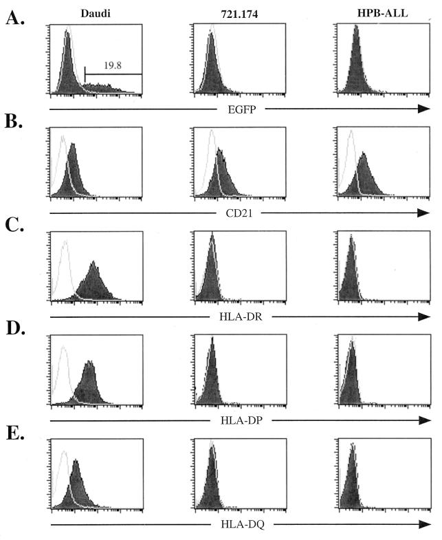
Susceptibility of CD21-expressing cell lines to EBfaV-GFP infection corresponds with surface expression of HLA class II molecules. (A) Daudi, 721.174, or HPB-ALL cells (106) were infected with 3 × 10⁵ green units as previously described (31). Twenty-four hours after infection, cells were analyzed for EGFP fluorescence by flow cytometry using a FacsCalibur (Becton Dickinson). Unshaded histograms represent EGFP fluorescence of cells in the absence of exposure to EBfaV-GFP. Shaded histograms represent EGFP fluorescence after exposure to the virus. The number above the marked region represents the percentage of Daudi cells infected. (B) Surface expression of CD21 on Daudi, 721.174, or HPB-ALL cells, analyzed by flow cytometry using the monoclonal antibody HB5 (34). Isotype-specific monoclonal antibodies were used to detect the surface expression of HLA-DR (TÜ36) (C), HLA-DP (HI43) (D), and HLA-DQ (Ia3) (E) on Daudi, 721.174, and HPB-ALL cells. Unshaded histograms (B to E) represent cells stained with an immunoglobulin isotype-matched control immunoglobulin G2a antibody recognized by a secondary goat anti-mouse antibody conjugated to allophycocyanin (Caltag Laboratories). Shaded histograms represent cells stained with the appropriate antibody recognized by the same secondary antibody.
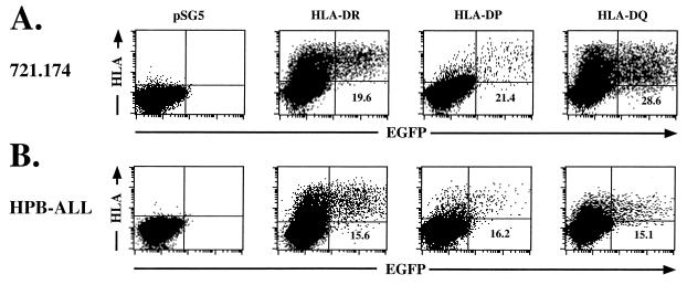
Expression of HLA-DR, -DP, or -DQ mediates EBV entry into 721.174 and HPB-ALL cells. Daudi or HPB-ALL cells (106) electroporated with HLA-DR, HLA-DP, or HLA-DQ were infected with EBfaV-GFP and analyzed by two-color flow cytometry. The flow cytometer was gated for EGFP fluorescence and class II expression using a pan-class II antibody, TÜ39, detected by a goat anti-mouse allophycocyanin-conjugated secondary antibody. pSG5-electroporated cells were exposed to EBfaV-GFP and stained identically to class II-transfected cells. The number given in the lower right quadrant represents the percentage of cells expressing the appropriate class II molecule which are infected by EBfaV-GFP. Each plot shows 40,000 events.
References
-
- Alkhatib G, Combadiere C, Broder C C, Feng Y, Kennedy P E, Murphy P M, Berger E A. CC CKR5: a RANTES, MIP-1alpha, MIP-1beta receptor as a fusion cofactor for macrophage-tropic HIV-1. Science. 1996;272:1955–1958. - PubMed
-
- Basham T, Smith W, Lanier L, Morhenn V, Merigan T. Regulation of expression of class II major histocompatibility antigens on human peripheral blood monocytes and Langerhans cells by interferon. Hum Immunol. 1984;10:83–93. - PubMed
-
- Basta P V, Sherman P A, Ting J P. Identification of an interferon-gamma response region 5′ of the human histocompatibility leukocyte antigen DR alpha chain gene which is active in human glioblastoma multiforme lines. J Immunol. 1987;138:1275–1280. - PubMed
Publication types
MeSH terms
Substances
Grants and funding
LinkOut - more resources
Full Text Sources
Other Literature Sources
Research Materials
