. 2000 Mar;74(5):2459-65.
doi: 10.1128/jvi.74.5.2459-2465.2000.

Adeno-associated virus major Rep78 protein disrupts binding of TATA-binding protein to the p97 promoter of human papillomavirus type 16

Affiliations

Adeno-associated virus major Rep78 protein disrupts binding of TATA-binding protein to the p97 promoter of human papillomavirus type 16

P F Su et al. J Virol. 2000 Mar.

Abstract

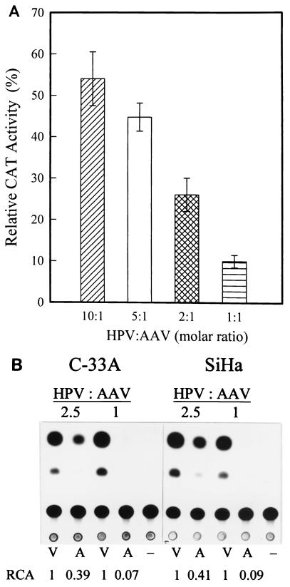
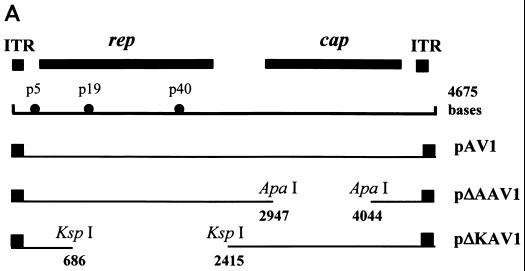
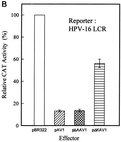
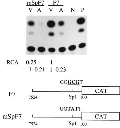
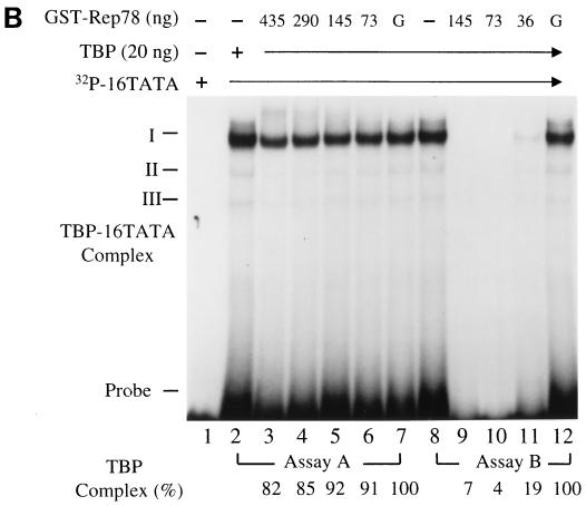
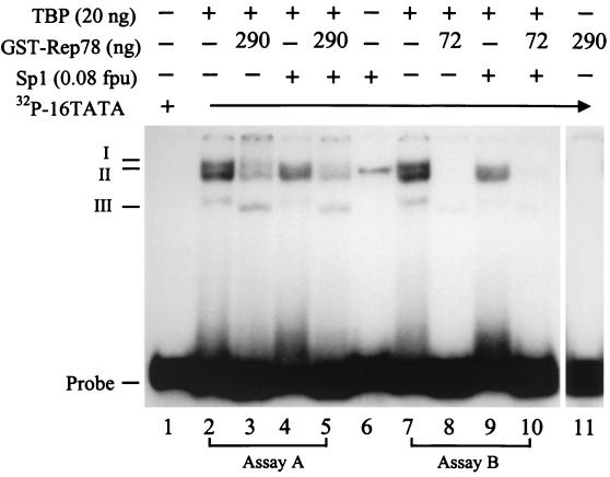
Adeno-associated virus type 2 (AAV) is known to inhibit the promoter activities of several oncogenes and viral genes, including the human papillomavirus type 16 (HPV-16) E6 and E7 transforming genes. However, the target elements of AAV on the long control region (LCR) upstream of E6 and E7 oncogenes are elusive. A chloramphenicol acetyltransferase assay was performed to study the effect of AAV on the transcription activity of the HPV-16 LCR in SiHa (HPV-positive) and C-33A (HPV-negative) cells. The results reveal that (i) AAV inhibited HPV-16 LCR activity in a dose-dependent manner, (ii) AAV-mediated inhibition did not require the HPV gene products, and (iii) the AAV replication gene product Rep78 was involved in the inhibition. Deletion mutation analyses of the HPV-16 LCR showed that regulatory elements outside the core promoter region of the LCR may not be direct targets of AAV-mediated inhibition. Further study with the electrophoretic mobility shift assay demonstrated that Rep78 interfered with the binding of TATA-binding protein (TBP) to the TATA box of the p97 core promoter more significantly than it disrupted the preformed TBP-TATA complex. These data thus suggest that Rep78 may inhibit transcription initiation of the HPV-16 LCR by disrupting the interaction between TBP and the TATA box of the p97 core promoter.


Figures

FIG. 1
FIG. 1
(A) Inhibitory effect of AAV on HPV-16 LCR promoter activity at different molar ratios. SiHa cells were cotransfected with pBL-16LCR-CAT6 reporter plasmid (10 μg) and either an AAV plasmid (pAV1, which carries WT AAV DNA within the pBR322 vector) (2, 4, 10, or 20 μg) or a control plasmid (pBR322; 1, 2, 5, or 10 μg) to yield HPV/AAV molar ratios of 10:1, 5:1, 2:1, and 1:1. Transfected cells were cultured for 48 h and harvested, and the cell extracts were prepared for CAT assay as described previously (25). Acetylated and nonacetylated spots were quantified by electronic autography with an instant imager (Packard, Meriden, Conn.). The promoter activity in extracts of cells cotransfected with pBL-16LCR-CAT6 and pBR322 was set as 100%. Calculation of the relative activity is described in text. The data are the means ± standard errors of four individual experiments. (B) Representative thin-layer chromatography analysis of the effect of AAV on the HPV-16 LCR in SiHa (HPV-positive) and C-33A (HPV-free) cells. The cells were cotransfected with reporter pBL-16LCR-CAT6 and either effector plasmid pAV1 or vector pBR3224 at molar ratios of 2.5:1 and 1:1. The subsequent steps were the same as described above. The chromatography results presented are from one of three individual experiments. −, pBLCAT6 (negative control); V, pBR322; A, pAV1; RCA, relative CAT activity.
FIG. 2
FIG. 2
Inhibitory effects of WT and mutant AAV genomes on activity of the HPV-16 LCR. (A) Plasmid constructs containing the WT or mutated AAV genome. ITR, inverted terminal repeat. (B) SiHa cells were cotransfected with 10 μg of reporter pBL-16LCR-CAT6 and either 5 μg of vector pBR322 or 10 μg of an effector plasmid (pAV1, pΔAAV1, or pΔKAV1) to yield a molar ratio of 2:1. The data are the means ± standard errors of four individual experiments.
FIG. 2
FIG. 2
Inhibitory effects of WT and mutant AAV genomes on activity of the HPV-16 LCR. (A) Plasmid constructs containing the WT or mutated AAV genome. ITR, inverted terminal repeat. (B) SiHa cells were cotransfected with 10 μg of reporter pBL-16LCR-CAT6 and either 5 μg of vector pBR322 or 10 μg of an effector plasmid (pAV1, pΔAAV1, or pΔKAV1) to yield a molar ratio of 2:1. The data are the means ± standard errors of four individual experiments.
FIG. 3
FIG. 3
Identification of motifs on the HPV-16 LCR involved in AAV-induced inhibition of promoter activity by mutation analyses. (A) Constructs of the WT and mutant (F1 to F7) HPV-16 LCR. The mutated LCR fragments were produced by PCR as outlined in Table 1 and were subsequently cloned into the vector pBLCAT6. (B) Promoter activities of WT and mutated HPV-16 LCR constructs. SiHa cells were cotransfected with 10 μg of reporter plasmid containing the WT or one of the mutated LCR constructs. The basal CAT activity from cells transfected with the WT construct was defined as 1, and the CAT activity from cells transfected with mutant LCR constructs is reported relative to that of the WT LCR. (C) Inhibitory effects of AAV on the promoter activities of WT and mutated HPV-16 LCR constructs. SiHa cells were cotransfected with 10 μg of a reporter construct carrying the WT or one of the truncated LCR fragments along with pAV1 or pBR322. The HPV/AAV molar ratio used for cotransfection was 2:1. pBLCAT6 and pSV2CAT were used as negative and positive controls, respectively (data not shown). −, not done. The results are reported as percentages of the value relative to the activity of samples from cells transfected with the same LCR construct plus pBR322. The data are the means ± standard errors of three individual experiments.
FIG. 4
FIG. 4
Inhibitory effects of AAV on F7 and mSpF7. SiHa cells were cotransfected with 10 μg of reporter plasmids containing F7 or mSpF7, along with pAV1 (A) or pBR322 vector (V). The molar ratio of HPV to AAV was 2. N and P, negative (pBLCAT6) and positive (pSV2CAT) controls, respectively. The data presented for relative CAT activity (RCA) are the averages of three individual experiments.
FIG. 5
FIG. 5
Effect of Rep78 on the complex formation of TBP and the HPV-16 p97 core promoter. (A) Sodium dodecyl sulfate-polyacrylamide gel electrophoresis and Coomassie blue staining of GST and GST-Rep78 proteins. The full-length AAV rep gene from nucleotide 321 to 2193 was amplified by PCR and cloned into vector pGEX-5X-3 (Pharmacia Biotech, Uppsala, Sweden) for expression of GST-Rep78 fusion protein. The protocol used for expression and purification of GST-Rep78 was as described by the manufacturer (Pharmacia). Lanes 1 and 3, GST-Rep78 and GST proteins eluted with 10 mM glutathione (reduced form); lanes 2 and 4, GST-Rep78 and GST protein conjugated on glutathione-Sepharose 4B beads. (B) EMSA analysis of the influence of GST-Rep78 on the interaction between TBP and the HPV-16 p97 TATA box. EMSA was performed in a total volume of 20 μl of reaction buffer containing 20 mM HEPES-KOH (pH 7.9), 25 mM KCl, 2 mM MgCl2, 0.1 mM EDTA, 0.5 mM dithiothreitol, 100 μg of bovine serum albumin per ml, 10% glycerol, and 0.025% Nonidet P-40. Twenty nanograms each of human recombinant TBP (Santa Cruz Biotechnology Inc., Santa Cruz, Calif.) and GST-Rep78 at the indicated concentrations was incubated with a 32P-labeled double-stranded 16TATA oligonucleotide (5′-AACGGTTAGTATAAAAGCAGACA), as described by Bauknecht and Shi (3), at 30°C for 30 min. The DNA-protein complexes were resolved on a 5% native polyacrylamide gel in 1× Tris-borate-EDTA (99 mM Tris base, 99 mM boric acid, and 2 mM EDTA [pH 8.3]) running buffer. Lane 1, free 32P-labeled 16TATA oligonucleotide; lanes 2 and 8, TBP-16TATA complexes; lanes 3 to 6, addition of GST-Rep78 to a reaction solution containing preformed TBP-16TATA complexes (assay A); lanes 9 to 11, incubation of TBP with GST-Rep78 prior to addition of 32P-labeled 16TATA oligonucleotide (assay B); lanes 7 and 12, addition of 435 and 145 ng of GST as a negative control for assays A and B, respectively. “G” represents GST and “Probe” represents a free 32P-labeled 16TATA oligonucleotide. The radioactivities of TBP-16TATA complexes on the gel were scored with an instant imager as described for Fig. 1. The percent TBP-16TATA complex formation was calculated as follows: (radioactivity of TBP-16TATA complex in the presence of GST-Rep78/radioactivity of TBP-16TATA complex in the presence of GST) × 100. I, II, and III, TBP-16 TATA complex.
FIG. 5
FIG. 5
Effect of Rep78 on the complex formation of TBP and the HPV-16 p97 core promoter. (A) Sodium dodecyl sulfate-polyacrylamide gel electrophoresis and Coomassie blue staining of GST and GST-Rep78 proteins. The full-length AAV rep gene from nucleotide 321 to 2193 was amplified by PCR and cloned into vector pGEX-5X-3 (Pharmacia Biotech, Uppsala, Sweden) for expression of GST-Rep78 fusion protein. The protocol used for expression and purification of GST-Rep78 was as described by the manufacturer (Pharmacia). Lanes 1 and 3, GST-Rep78 and GST proteins eluted with 10 mM glutathione (reduced form); lanes 2 and 4, GST-Rep78 and GST protein conjugated on glutathione-Sepharose 4B beads. (B) EMSA analysis of the influence of GST-Rep78 on the interaction between TBP and the HPV-16 p97 TATA box. EMSA was performed in a total volume of 20 μl of reaction buffer containing 20 mM HEPES-KOH (pH 7.9), 25 mM KCl, 2 mM MgCl2, 0.1 mM EDTA, 0.5 mM dithiothreitol, 100 μg of bovine serum albumin per ml, 10% glycerol, and 0.025% Nonidet P-40. Twenty nanograms each of human recombinant TBP (Santa Cruz Biotechnology Inc., Santa Cruz, Calif.) and GST-Rep78 at the indicated concentrations was incubated with a 32P-labeled double-stranded 16TATA oligonucleotide (5′-AACGGTTAGTATAAAAGCAGACA), as described by Bauknecht and Shi (3), at 30°C for 30 min. The DNA-protein complexes were resolved on a 5% native polyacrylamide gel in 1× Tris-borate-EDTA (99 mM Tris base, 99 mM boric acid, and 2 mM EDTA [pH 8.3]) running buffer. Lane 1, free 32P-labeled 16TATA oligonucleotide; lanes 2 and 8, TBP-16TATA complexes; lanes 3 to 6, addition of GST-Rep78 to a reaction solution containing preformed TBP-16TATA complexes (assay A); lanes 9 to 11, incubation of TBP with GST-Rep78 prior to addition of 32P-labeled 16TATA oligonucleotide (assay B); lanes 7 and 12, addition of 435 and 145 ng of GST as a negative control for assays A and B, respectively. “G” represents GST and “Probe” represents a free 32P-labeled 16TATA oligonucleotide. The radioactivities of TBP-16TATA complexes on the gel were scored with an instant imager as described for Fig. 1. The percent TBP-16TATA complex formation was calculated as follows: (radioactivity of TBP-16TATA complex in the presence of GST-Rep78/radioactivity of TBP-16TATA complex in the presence of GST) × 100. I, II, and III, TBP-16 TATA complex.
FIG. 6
FIG. 6
EMSA analysis of the effect of Rep78 on the interaction of TBP and the 16TATA oligonucleotide in the presence of Sp1. EMSA was performed as described for Fig. 5B except that 0.5× TBE running buffer was used. In assay A, GST-Rep78 and Sp1 (Promega Corporation, Madison, Wis.) were added to preformed TBP-16TATA complex individually or mixed (lanes 3 to 5). In assay B, TBP was incubated with GST-Rep78 and Sp1 separately or together prior to the addition of 32P-labeled 16TATA oligonucleotide (lanes 8 to 10). Lane 1, free 32P-labeled 16TATA oligonucleotide; lanes 2 and 7, TBP-16TATA complex; lane 3, addition of GST-Rep78 to the TBP-16TATA complex; lane 4, addition of Sp1 to the TBP-16TATA complex; lane 5, addition of a mixture of GST-Rep78 and Sp1 to the TBP-16TATA complex; lane 6, the Sp1-16TATA complex; lane 8, incubation of TBP with GST-Rep78 prior to the addition of 16TATA oligonucleotide; lane 9, incubation of TBP and Sp1 prior to addition of the 16TATA oligonucleotide; lane 10, incubation of TBP with a mixture of GST-Rep78 and Sp1 before the addition of the 16TATA oligonucleotide; lane 11, incubation of GST-Rep78 with 16TATA probe as a negative control. “Probe” indicates a free 32P-labeled 16TATA oligonucleotide. I, II, and III, TBP-16TATA complex; fpu, footprinting unit.

Similar articles

Cited by

References

    1. Antoni B A, Rabson A B, Miller I L, Trempe J P, Chejanovsky N, Carter B J. Adeno-associated virus Rep protein inhibits human immunodeficiency virus type 1 production in human cells. J Virol. 1991;65:396–404. - PMC - PubMed
    1. Apt D, Watts R M, Suske G, Bernard H U. High Sp1/Sp3 ratios in epithelial cells during epithelial differentiation and cellular transformation correlate with the activation of the HPV-16 promoter. Virology. 1996;224:281–291. - PubMed
    1. Bauknecht T, Shi Y. Overexpression of C/EBPβ represses human papillomavirus type 18 upstream regulatory region activity in HeLa cells by interfering with the binding of TATA-binding protein. J Virol. 1998;72:2113–2124. - PMC - PubMed
    1. Beaton A, Palumbo P, Berns K I. Expression from the adeno-associated virus p5 and p19 promoters is negatively regulated in trans by the rep protein. J Virol. 1989;63:4450–4454. - PMC - PubMed
    1. Berns K I. Parvoviridae: the viruses and their replication. In: Fields B N, Knipe D M, Howley P M, editors. Fields virology. 3rd ed. Vol. 2. Philadelphia, Pa: Lippincott-Raven; 1996. pp. 2173–2197.

Publication types

LinkOut - more resources