. 2000 Feb;156(2):393-8.
doi: 10.1016/S0002-9440(10)64742-6.

Control of apoptosis during angiogenesis by survivin expression in endothelial cells

Affiliations

Control of apoptosis during angiogenesis by survivin expression in endothelial cells

D S O'Connor et al. Am J Pathol. 2000 Feb.

Abstract

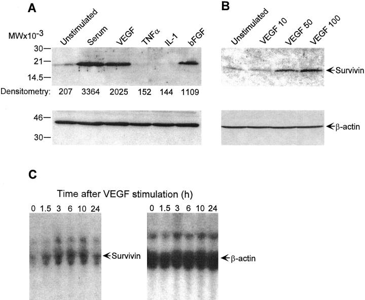
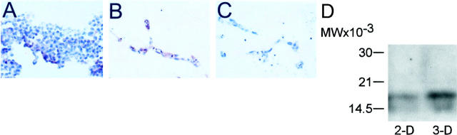
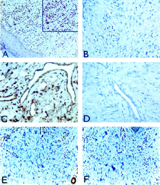
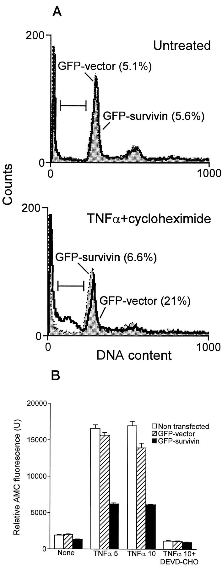
Mechanisms controlling endothelial cell survival during angiogenesis were investigated. Stimulation of quiescent endothelial cells with mitogens, including vascular endothelial growth factor and basic fibroblast growth factor, induced up to approximately 16-fold up-regulation of the cell cycle-regulated apoptosis inhibitor survivin. Mitogen stimulation rapidly increased survivin RNA expression in endothelial cells, which peaked after 6 to 10 hours in culture and decreased by 24 hours. Inflammatory cytokines, tumor necrosis factor alpha, and interleukin-1 did not induce survivin expression in endothelial cells. Formation of three-dimensional vascular tubes in vitro was associated with strong induction of survivin in endothelial cells, as compared with two-dimensional cultures. By immunohistochemistry, survivin was minimally expressed in endothelium of nonproliferating capillaries of normal skin, whereas it became massively up-regulated in newly formed blood vessels of granulation tissue in vivo. Recombinant expression of green fluorescent protein survivin in endothelial cells reduced caspase-3 activity and counteracted apoptosis induced by tumor necrosis factor alpha/cycloheximide. These findings identify survivin as a novel growth factor-inducible protective gene expressed by endothelial cells during angiogenesis. Therapeutic manipulation of survivin expression and function in endothelium may influence compensatory or pathological (tumor) angiogenesis.


Figures

Figure 1.
Figure 1.
Modulation of survivin expression in EC. A: Quiescent EC were incubated with medium or serum (10% FCS), VEGF (100 ng/ml), bFGF (5 ng/ml), TNFα (10 ng/ml), or IL-1 (2 ng/ml) for 16 hours at 37°C. Cells were harvested, sodium dodecyl sulfate-extracted, and analyzed for expression of survivin or β-actin by immunoblotting. B: Control or EC were stimulated with the indicated increasing concentrations of VEGF for 16 hours at 37°C and analyzed for expression of survivin or β-actin by immunoblotting. C: Total RNA was extracted from EC stimulated with 100 ng/ml VEGF at the indicated time intervals, separated on agarose-formaldehyde denaturing gels, and hybridized with probes to survivin or control β-actin.
Figure 2.
Figure 2.
Expression of survivin in three-dimensional EC culture. EC were grown in three-dimensional fibronectin-collagen gels, paraffin-embedded, and analyzed for survivin expression (red staining) by immunohistochemistry. A: Survivin expression in control, two-dimensional EC culture. B: Survivin expression in three-dimensional EC culture. C: Control staining of three-dimensional EC culture with preimmune antibody. D: Two- (2-D) or three-dimensional (3-D) EC cultures were harvested, homogenized in a tissue grinder, and analyzed for survivin expression by immunoblotting.
Figure 3.
Figure 3.
Expression of survivin in proliferating and nonproliferating skin capillaries. Five-micron sections of formalin-fixed, paraffin-embedded skin biopsies containing granulation tissue and normal skin were analyzed for survivin expression by immunohistochemistry after antigen retrieval by pressure-cooking. A: Strong cytoplasmic expression of survivin in EC of dermal capillaries in granulation tissue. Inset: Detail representation of dermal capillaries stained for survivin expression. C: Expression of survivin in endothelium of large vessel in granulation tissue at the dermis/hypodermis junction. B and D: Control staining for A and C, respectively, in the absence of primary antibody. E: Expression of survivin in nonproliferating capillaries of noninflamed normal skin. F: Control incubation for panel E in the presence of preimmune antibody. Original magnifications, ×200 (A, E, F) and ×400 (B, C, D).
Figure 4.
Figure 4.
Anti-apoptotic function of survivin in EC. A: Subconfluent bovine aortic EC were transfected with GFP vector or GFP survivin by lipofectin, cultivated for 35 hours at 37°C, and treated with 5 ng/ml TNFα/5 μg/ml cycloheximide for additional 8 hours at 37°C. GFP-expressing cells (green fluorescence) were analyzed for DNA content by propidium iodide staining (red fluorescence) and flow cytometry. The percentage of cells with hypodiploid DNA content (sub-G1 fraction) is indicated in parentheses for each condition tested. B: EC, untreated or transfected with GFP vector or GFP survivin, were incubated with the indicated concentrations of TNFα/10 μg/ml cycloheximide, harvested, and analyzed for caspase-3 activity by hydrolysis of the fluorogenic substrate DEVD-AMC in the presence or absence of the caspase-3 inhibitor DEVD-CHO. Data are the mean ± SD of replicates of a representative experiment.

References

    1. Vaux DL, Korsmeyer SJ: Cell death in development. Cell 1999, 96:245-254 - PubMed
    1. Salvesen GS, Dixit VM: Caspases: intracellular signaling by proteolysis. Cell 1997, 91:443-446 - PubMed
    1. Thompson CB: Apoptosis in the pathogenesis and treatment of disease. Science 1995, 267:1456-1462 - PubMed
    1. Rudin CM, Thompson CB: Apoptosis and disease: regulation and clinical relevance of programmed cell death. Annu Rev Med 1997, 48:267-281 - PubMed
    1. Bjorkerud S, Bjorkarud B: Apoptosis is abundant in human atherosclerotic lesions, especially in inflammatory cells (macrophages and T cells), and may contribute to the accumulation of gruel and plaque instability. Am J Pathol 1996, 149:367-380 - PMC - PubMed

Publication types

MeSH terms