. 2000 Feb;156(2):545-53.
doi: 10.1016/S0002-9440(10)64759-1.

Localization of cyclooxygenase-2 in human sporadic colorectal adenomas

Affiliations

Localization of cyclooxygenase-2 in human sporadic colorectal adenomas

K S Chapple et al. Am J Pathol. 2000 Feb.

Abstract

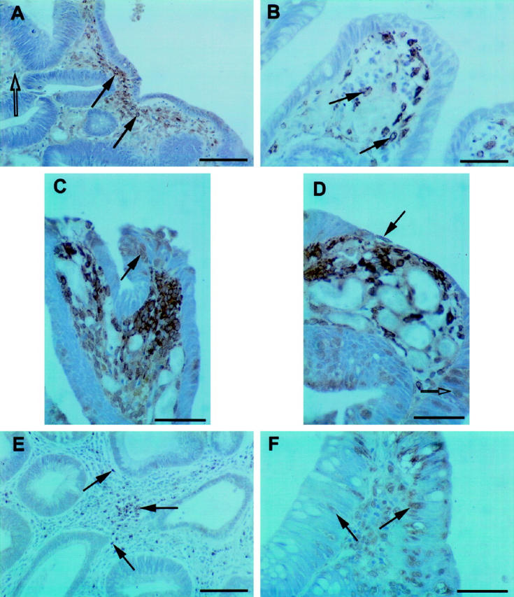
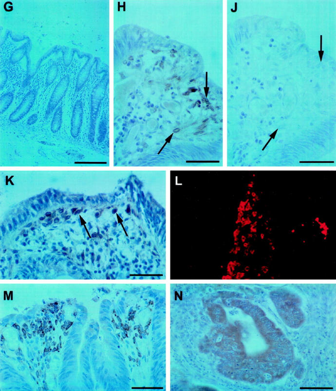
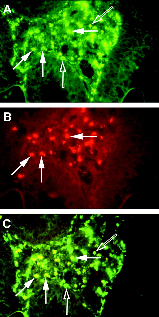
A putative target for the anti-colorectal cancer action of nonsteroidal anti-inflammatory drugs is the inducible isoform of cyclooxygenase (COX), COX-2. COX-2 is expressed within intestinal adenomas in murine polyposis models, but expression has been poorly characterized in human colorectal neoplasms. Therefore, we investigated the localization of the COX-2 protein in human sporadic colorectal adenomas. Immunohistochemistry for COX-2 and CD68 (a tissue macrophage marker) was performed on formalin-fixed, paraffin-embedded (n = 52) and frozen, acetone-fixed (n = 6) sections of human sporadic colorectal adenomas. Forty of 52 (77%) formalin-fixed adenomas expressed immunoreactive COX-2. COX-2 was localized to superficial interstitial macrophages in 39 cases (75%) and to deep interstitial macrophages in 9 cases (17%). COX-2 staining of dysplastic epithelial cells was observed in 15 cases (29%). A logistic regression analysis identified the adenoma site (P = 0.012) and histological type (P = 0.001) as independent predictors of superficial macrophage COX-2 expression. There was no relationship between the number of macrophages within an adenoma and macrophage COX-2 expression. These results indicate that COX-2 is expressed predominantly by interstitial macrophages within human sporadic colorectal adenomas. If COX-2 does indeed play a role in the early stages of colorectal carcinogenesis in man, these data suggest COX-2-mediated paracrine signaling between the macrophages and epithelial cells within adenomas.


Figures

Figure 1.
Figure 1.
Immunohistochemistry for COX-2 on human sporadic colorectal adenomas. Affinity-purified rabbit polyclonal anti-human COX-2 IgG was used unless stated otherwise. A: COX-2 immunostaining of superficial interstitial cells (closed arrows) adjacent to COX-2-negative epithelial cells at the flat luminal surface of an adenoma. Within adenomas, aggregates of COX-2-positive superficial interstitial cells were separated by interstitium containing no COX-2-positive cells (open arrow). Scale bar = 100 μm. B: COX-2-positive superficial interstitial cells (arrows) at the tip of a dysplastic villus. Cells were mononuclear with large ovoid nuclei. COX-2 immunoreactivity was demonstrated in a perinuclear distribution (arrows; see also panels C, D). Scale bar = 50 μm. C: Numerous COX-2-positive superficial interstitial cells in close proximity to capillaries (see also D). The epithelium contained occasional COX-2-positive cells (arrow; see also D, F). Scale bar = 50 μm. D: COX-2-positive cells adjacent to flattened epithelium (closed arrow) at the tip of a villus. Note the densely vascular area immediately adjacent to the COX-2-positive cell infiltrate. COX-2-positive epithelial cells were also observed (open arrow). Scale bar = 50 μm. E: COX-2-positive interstitial cells deep within an adenoma (arrows). In contrast to superficial interstitial cells (see A–D), deep interstitial cells were smaller and less closely aggregated. Scale bar = 100 μm. F: COX-2 localization to epithelial cells (arrows). Staining was abolished by antibody preadsorption with its cognate peptide. Scale bar = 50 μm. G: Normal colonic mucosa. No COX-2 immunoreactivity was detected. Scale bar = 200 μm. H: COX-2 localization to superficial interstitial cells (arrows). Scale bar = 50 μm. J: Preadsorption of COX-2 antibody with its cognate peptide. There was abolition of specific COX-2 staining compared with panel H (arrows). Scale bar = 50 μm. K: COX-2 immunohistochemistry on frozen, acetone-fixed tissue, using peroxidase detection. Localization of COX-2 to superficial interstitial cells (arrows) was similar to that demonstrated in fixed tissue (compare with A) and was abolished by antibody preincubation with its cognate peptide. Faint staining of the epithelium was non-specific and was not abolished by antibody pre-adsorption. Scale bar = 50 μm. L: COX-2 immunohistochemistry on frozen, acetone-fixed tissue, using fluorescent detection (TRITC). Numerous COX-2-positive interstitial cells (red) were observed. No epithelial cell staining was detected. M: COX-2 immunohistochemistry on formalin-fixed, paraffin-embedded sections, using murine COX-2 antiserum. There was a similar distribution of immunoreactivity to that obtained using rabbit polyclonal anti-human COX-2 IgG (see A−D). Scale bar = 50 μm. N: Human sporadic colorectal carcinoma. Diffuse cytoplasmic COX-2 staining was apparent in cancer cells. Scale bar = 50 μm.
Figure 1.
Figure 1.
Immunohistochemistry for COX-2 on human sporadic colorectal adenomas. Affinity-purified rabbit polyclonal anti-human COX-2 IgG was used unless stated otherwise. A: COX-2 immunostaining of superficial interstitial cells (closed arrows) adjacent to COX-2-negative epithelial cells at the flat luminal surface of an adenoma. Within adenomas, aggregates of COX-2-positive superficial interstitial cells were separated by interstitium containing no COX-2-positive cells (open arrow). Scale bar = 100 μm. B: COX-2-positive superficial interstitial cells (arrows) at the tip of a dysplastic villus. Cells were mononuclear with large ovoid nuclei. COX-2 immunoreactivity was demonstrated in a perinuclear distribution (arrows; see also panels C, D). Scale bar = 50 μm. C: Numerous COX-2-positive superficial interstitial cells in close proximity to capillaries (see also D). The epithelium contained occasional COX-2-positive cells (arrow; see also D, F). Scale bar = 50 μm. D: COX-2-positive cells adjacent to flattened epithelium (closed arrow) at the tip of a villus. Note the densely vascular area immediately adjacent to the COX-2-positive cell infiltrate. COX-2-positive epithelial cells were also observed (open arrow). Scale bar = 50 μm. E: COX-2-positive interstitial cells deep within an adenoma (arrows). In contrast to superficial interstitial cells (see A–D), deep interstitial cells were smaller and less closely aggregated. Scale bar = 100 μm. F: COX-2 localization to epithelial cells (arrows). Staining was abolished by antibody preadsorption with its cognate peptide. Scale bar = 50 μm. G: Normal colonic mucosa. No COX-2 immunoreactivity was detected. Scale bar = 200 μm. H: COX-2 localization to superficial interstitial cells (arrows). Scale bar = 50 μm. J: Preadsorption of COX-2 antibody with its cognate peptide. There was abolition of specific COX-2 staining compared with panel H (arrows). Scale bar = 50 μm. K: COX-2 immunohistochemistry on frozen, acetone-fixed tissue, using peroxidase detection. Localization of COX-2 to superficial interstitial cells (arrows) was similar to that demonstrated in fixed tissue (compare with A) and was abolished by antibody preincubation with its cognate peptide. Faint staining of the epithelium was non-specific and was not abolished by antibody pre-adsorption. Scale bar = 50 μm. L: COX-2 immunohistochemistry on frozen, acetone-fixed tissue, using fluorescent detection (TRITC). Numerous COX-2-positive interstitial cells (red) were observed. No epithelial cell staining was detected. M: COX-2 immunohistochemistry on formalin-fixed, paraffin-embedded sections, using murine COX-2 antiserum. There was a similar distribution of immunoreactivity to that obtained using rabbit polyclonal anti-human COX-2 IgG (see A−D). Scale bar = 50 μm. N: Human sporadic colorectal carcinoma. Diffuse cytoplasmic COX-2 staining was apparent in cancer cells. Scale bar = 50 μm.
Figure 2.
Figure 2.
Colocalization of COX-2 and CD68 in human sporadic colorectal adenoma by dual labeling immunofluorescence. A: Numerous CD68-positive macrophages (green; open and closed arrows). B: COX-2-positive interstitial cells (red; arrows). C: Colocalization of COX-2 and CD68 using a dual wavelength filter. Closed arrows indicate COX-2-positive, CD68-positive macrophages (yellow), open arrows indicate COX-2-negative, CD68-positive macrophages (green).

Similar articles

Cited by

References

    1. Levy GN: Prostaglandin H synthases, nonsteroidal anti-inflammatory drugs, and colon cancer. FASEB J 1997, 11:234-247 - PubMed
    1. Non-steroidal anti-inflammatory drugs. IARC Handbooks of Cancer Prevention, vol 1. Lyon, France, International Agency for Research on Cancer, 1997
    1. Shiff SJ, Rigas B: Nonsteroidal anti-inflammatory drugs and colorectal cancer: evolving concepts of their chemopreventive actions. Gastroenterology 1997, 113:1992-1998 - PubMed
    1. Herschman HR: Prostaglandin synthase 2. Biochim Biophys Acta 1996, 1299:125-140 - PubMed
    1. Sano H, Kawahito Y, Wilder RL, Hashiramoto A, Mukai S, Asai K, Kimura S, Kato H, Kondo M, Hla T: Expression of cyclooxygenase-1 and -2 in human colorectal cancer. Cancer Res 1995, 55:3785-3789 - PubMed

Publication types