

doi: 10.1093/nar/28.5.1067.
A simple in vitro Tn7-based transposition system with low target site selectivity for genome and gene analysis
Affiliations
- PMID: 10666445
- PMCID: PMC102592
- DOI: 10.1093/nar/28.5.1067
Item in Clipboard
A simple in vitro Tn7-based transposition system with low target site selectivity for genome and gene analysis
Nucleic Acids Res.
.
Abstract
A robust Tn7-based in vitro transposition system is described that displays little target site selectivity, allowing the efficient recovery of many different transposon insertions in target DNAs ranging from small plasmids to cosmids to whole genomes. Two miniTn7 derivatives are described that are useful for the analysis of genes: one a derivative for making translational and transcriptional target gene fusions and the other a derivative that can generate 15 bp (5 amino acid) insertions in target DNAs (proteins).
Figures
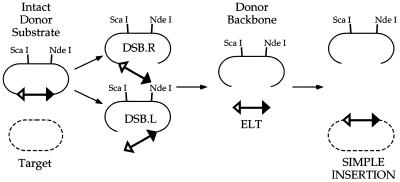
Substrates, reaction intermediates and products of Tn7 transposition. The donor molecule is disconnected from the donor backbone by DSBs at the ends of Tn7, generating Double-Strand Break Left (DSB.L) and Double-Strand Break Right (DSB.R) intermediates and an excised linear transposon (ELT); the ELT is joined to the target DNA to generate a simple insertion. Target DNA is represented by broken lines, donor DNA by solid lines. The filled triangle represents the right end of Tn7 and the open triangle, the left end. NdeI and ScaI sites are indicated in the donor backbone; NdeI was used to linearize pEMΔ and ScaI linearizes the other donors used.
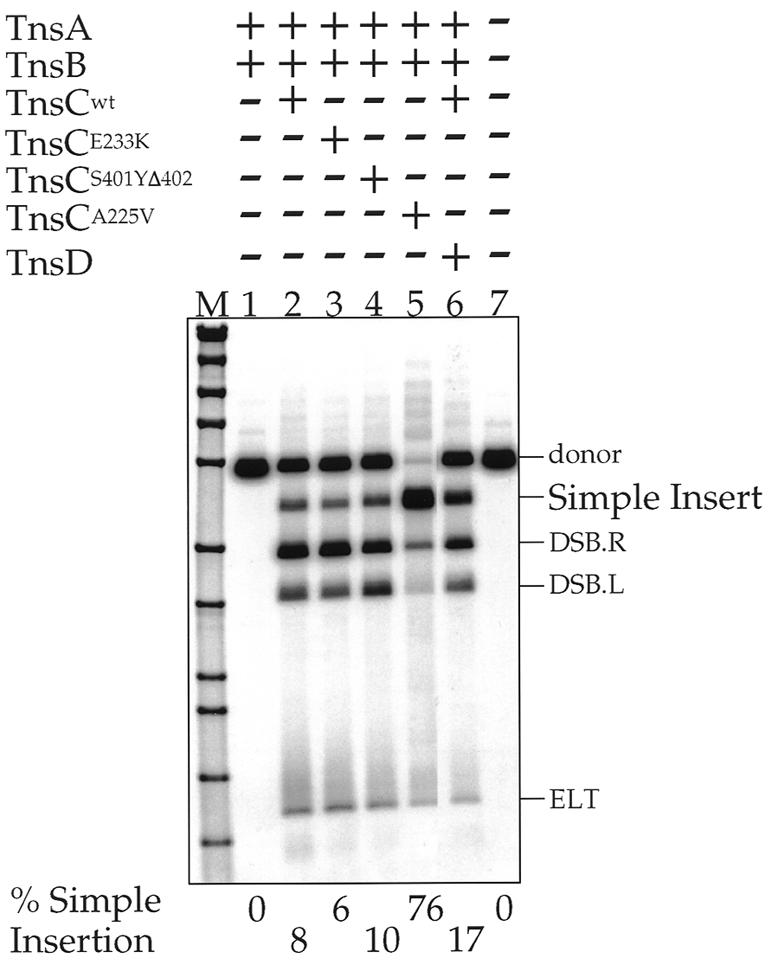
A Southern blot of NdeI-digested in vitro transposition products resolved on an agarose gel, transferred to a membrane and detected by using a transposon-specific probe. TnsABCmut reactions are compared to the TnsABCwt and TnsABC+D reactions; the percentage of donor substrate converted to simple insertion is shown beneath each lane. The various DNAs are labeled as described in Figure 1.
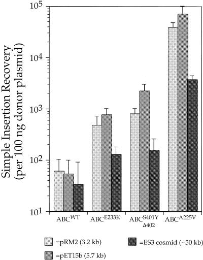
Histograms showing the recovery of transposition products via transformation of simple insertion products from in vitro TnsABCmut and TnsABCwt transposition reactions. The donor plasmid used was pMCB40 (p-oriγR6K:: miniTn7 SpeI–KmR–NotI) which cannot replicate in the transformed strain. Three differently sized target plasmids, pRM2, pET15b and ES3 were used.
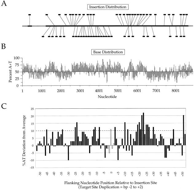
(A) Distribution of 63 insertions generated by TnsABCA225V in vitro transposition of miniTn7 from pEMΔ into target plasmid pER183, recovered by transformation and analyzed by DNA sequencing; junction sequences were determined utilizing Tn7 end-specific sequencing primers for both ends. Left→right insertions are indicated on the top, and right→left insertions are on the bottom. Essential regions of the target excluded by selection are indicated in gray. (B) Base composition of pER183, presented as percentage A+T for each 20 bp along the length of the target sequence. (C) Diagram of the nucleotide sequences flanking the 63 positions of Tn7 insertion in pER183, centered on the 5 bp target duplication. The vertical axis denotes the percentage by which the A+T content for a given position deviates from the average A+T content of the entire plasmid. The horizontal axis represents nucleotide position relative to the 5 bp target sequence duplication, with positions to the left of the insertion site indicated as negative numbers and positions to the right as positive numbers.
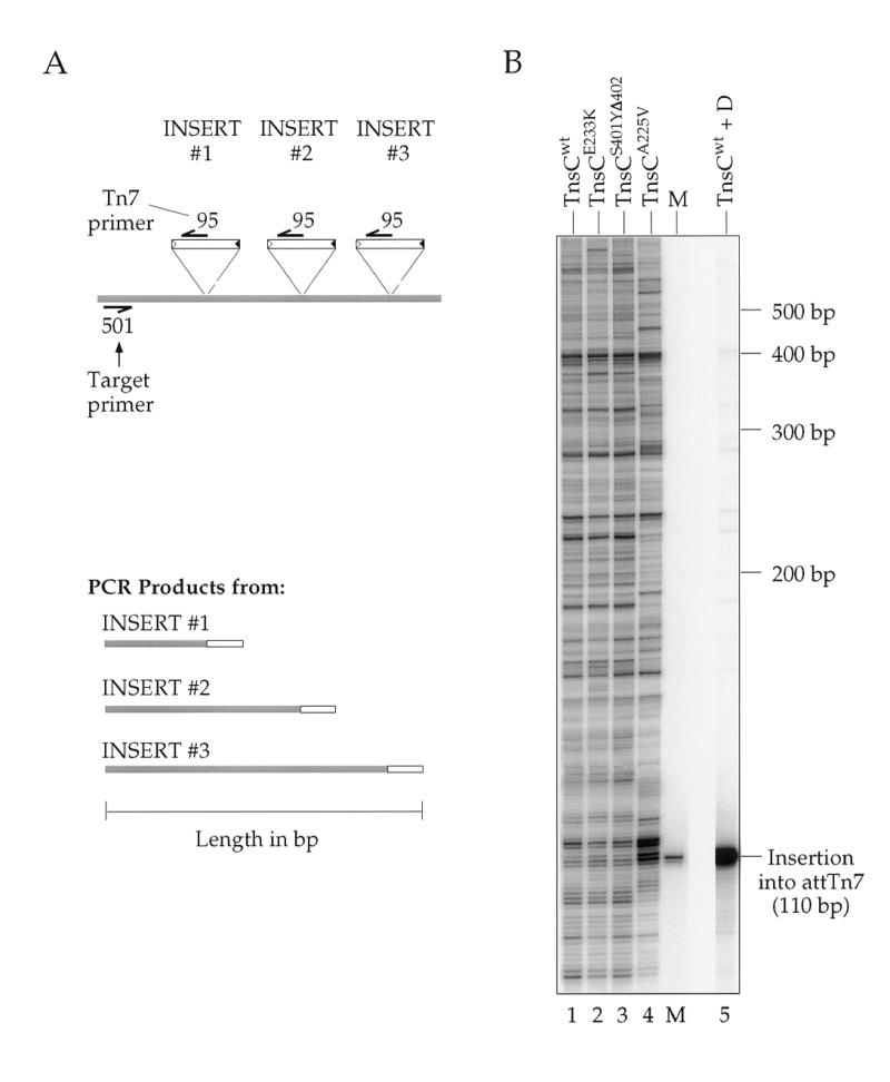
Analysis by a Southern blot of PCR products amplified from in vitro reactions using different TnsCs resolved on a denaturing gel and hybridized to a transposon-specific probe. (A) Diagram depicting three independent insertions which might occur in a given transposition reaction, and the corresponding products that would arise from PCR amplification with target-specific primer NLC501 and Tn7 end-specific primer NLC95. The shaded regions indicate target DNA or the portion of DNA in a PCR product that is derived from target template. (B) The profiles for three gain-of-function mutant TnsC reactions are compared to those for TnsABCwt and TnsABC+D in target plasmid pRM2. The amplicon chosen encompasses the attTn7 insertion site, allowing observation of the strong preference the TnsABC+D reaction (lane 5) displays for this position. The marker (M) is a PCR product amplified from an isolated insertion into attTn7 via a TnsABC+D reaction.
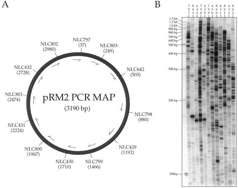
(A) Schematic illustrating the positions of 12 different target-specific PCR primers used to survey the positions of TnsABC225V-mediated insertions into into pRM2. (B) Southern blots of PCR products derived from amplification of transposition reactions using the indicated target primers and hybridized to end-labeled oligonucleotides, NLC646 and NLC685.
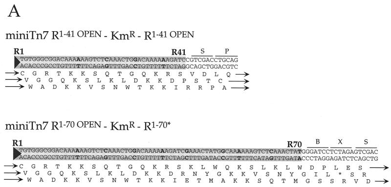
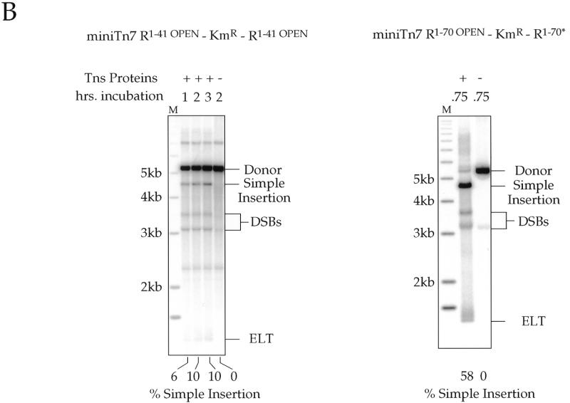
(A) Sequence of the Tn7 ends in two miniTn7 elements: nucleotide substitutions made to wild-type Tn7R end sequences are indicated in bold. Diagrams of the termini of the donor plasmids containing Tn7 termini that have open reading frames through them; the amino acid translation is shown beneath each end sequence, with ‘*’ indicating stop codons. B, BamHI site; X, XbaI site; S, SalI site; P, PstI site. These read-through elements are R1–41 OPEN–KmR–R1–41 OPEN (pMCB62) and R1–70 OPEN–KmR–R1–70* (pMCB64). R70* is a partially mutated right end that is mutated as in the R1–41 OPEN element and wild-type from R42–R70. (B) Southern blots of ScaI-digested transposition products of in vitro TnsABCA225V reactions for the two donor plasmids in (A), as detected by a hybridization with a transposon specific probe; the target plasmid for these experiments was pRM2. The reactions were incubated for various times as indicated. About 10% of R1–41 OPEN–KmR–R1–41 OPEN was converted to simple insertions; ∼58% of R1–70 OPEN–KmR–R1–70* was converted to simple insertion. The DNA species are labeled as in Figure 1.
(A) Sequence of the Tn7 ends in two miniTn7 elements: nucleotide substitutions made to wild-type Tn7R end sequences are indicated in bold. Diagrams of the termini of the donor plasmids containing Tn7 termini that have open reading frames through them; the amino acid translation is shown beneath each end sequence, with ‘*’ indicating stop codons. B, BamHI site; X, XbaI site; S, SalI site; P, PstI site. These read-through elements are R1–41 OPEN–KmR–R1–41 OPEN (pMCB62) and R1–70 OPEN–KmR–R1–70* (pMCB64). R70* is a partially mutated right end that is mutated as in the R1–41 OPEN element and wild-type from R42–R70. (B) Southern blots of ScaI-digested transposition products of in vitro TnsABCA225V reactions for the two donor plasmids in (A), as detected by a hybridization with a transposon specific probe; the target plasmid for these experiments was pRM2. The reactions were incubated for various times as indicated. About 10% of R1–41 OPEN–KmR–R1–41 OPEN was converted to simple insertions; ∼58% of R1–70 OPEN–KmR–R1–70* was converted to simple insertion. The DNA species are labeled as in Figure 1.
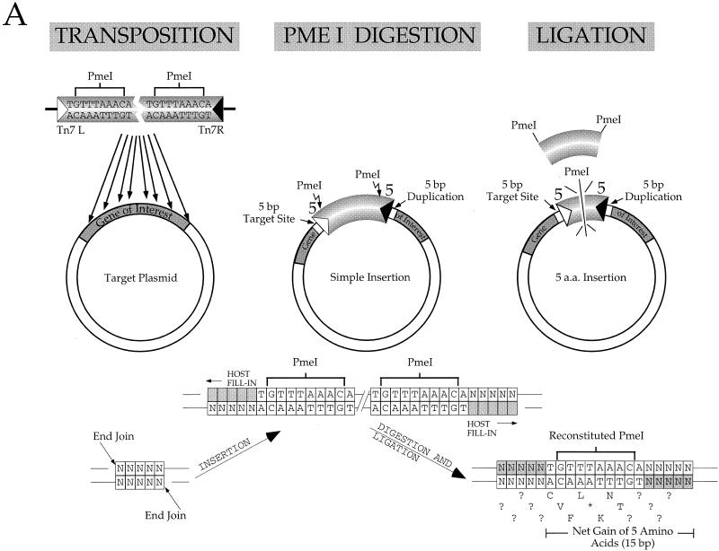
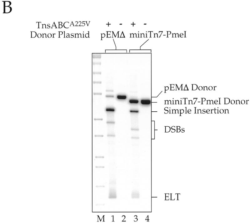
(A) Schematic diagram for making a 5 amino acid (15 bp) insertion in a target gene. A miniTn7 element containing PmeI sites close to the transposon termini is inserted into the target DNA at many different sites by transposition. The target plasmid with its inserted miniTn7 element DNA is then isolated, digested with PmeI and religated, resulting in a 15 bp insertion in the target DNA, 5 bp from each end of Tn7 and 5 bp from the target site duplication. Some of the inserted amino acids are dictated by the Tn7 end sequences whereas others will vary due to variations in the target sequence. (B) Southern blot using a transposon-specific probe of restriction digested products of in vitro transposition reactions using miniTn7 PmeI as a donor; pRM2 was the target DNA. Reactions containing pEMΔ were digested with NdeI and those using miniTn7 PmeI were digested with ScaI. DNA species are labeled as in Figure 1.
(A) Schematic diagram for making a 5 amino acid (15 bp) insertion in a target gene. A miniTn7 element containing PmeI sites close to the transposon termini is inserted into the target DNA at many different sites by transposition. The target plasmid with its inserted miniTn7 element DNA is then isolated, digested with PmeI and religated, resulting in a 15 bp insertion in the target DNA, 5 bp from each end of Tn7 and 5 bp from the target site duplication. Some of the inserted amino acids are dictated by the Tn7 end sequences whereas others will vary due to variations in the target sequence. (B) Southern blot using a transposon-specific probe of restriction digested products of in vitro transposition reactions using miniTn7 PmeI as a donor; pRM2 was the target DNA. Reactions containing pEMΔ were digested with NdeI and those using miniTn7 PmeI were digested with ScaI. DNA species are labeled as in Figure 1.
References
-
- Kleckner N., Bender,J. and Gottesman,S. (1991) Methods Enzymol., 204, 139–180. - PubMed
-
- Kleckner N., Roth,J. and Botstein,D. (1977) J. Mol. Biol., 116, 125–159. - PubMed
-
- Kaiser K., Sentry,J.W. and Finnegan,D.J. (1995) In Sherratt,D.J. (ed.), Eukaryotic Transposable Elements as Tools to Study Gene Structure and Function. IRL Press, Oxford, pp. 69–100.
-
- Berg C.M. and Berg,E. (1995) In Sherratt,D.J. (ed.), Transposable Elements as Tools for Molecular Analyses in Bacteria. IRL Press, Oxford, pp. 38–68.
-
- Waddell C.S. and Craig,N.L. (1988) Genes Dev., 2, 137–149. - PubMed
Publication types
MeSH terms
Substances
Grants and funding
LinkOut - more resources
Full Text Sources
Other Literature Sources
