. 2000 Mar 1;28(5):1106-13.
doi: 10.1093/nar/28.5.1106.

Transactivation and growth suppression by the gut-enriched Krüppel-like factor (Krüppel-like factor 4) are dependent on acidic amino acid residues and protein-protein interaction

Affiliations

Transactivation and growth suppression by the gut-enriched Krüppel-like factor (Krüppel-like factor 4) are dependent on acidic amino acid residues and protein-protein interaction

D E Geiman et al. Nucleic Acids Res. .

Abstract

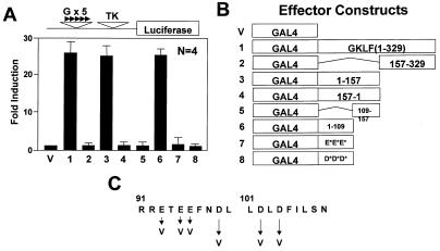
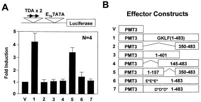
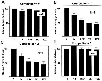
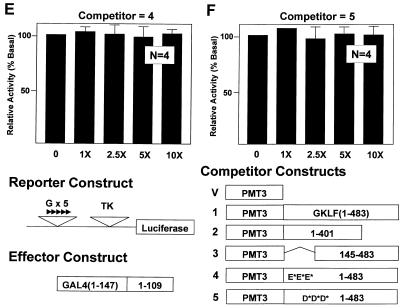
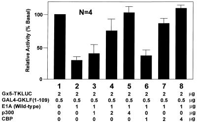
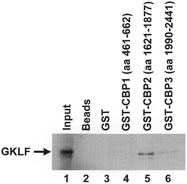
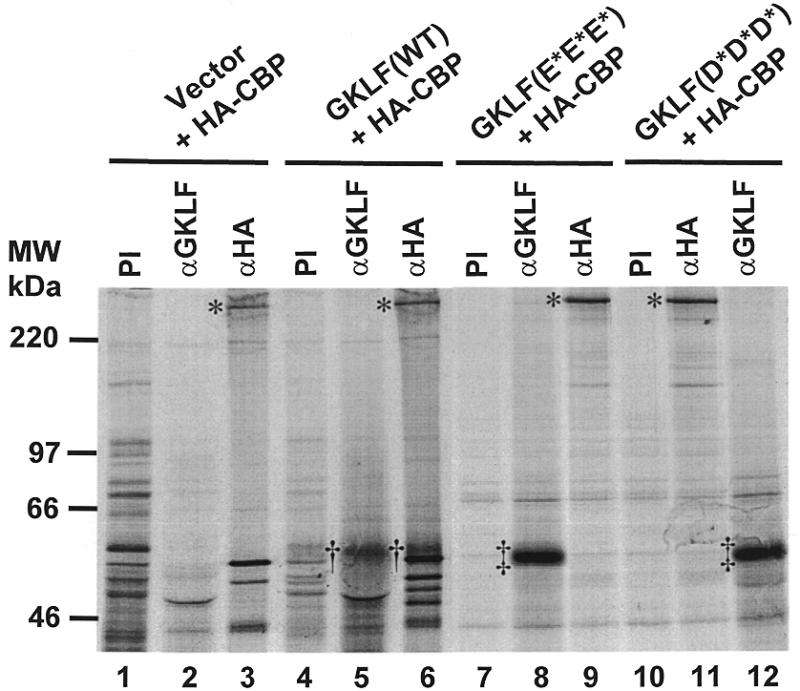
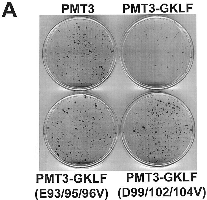
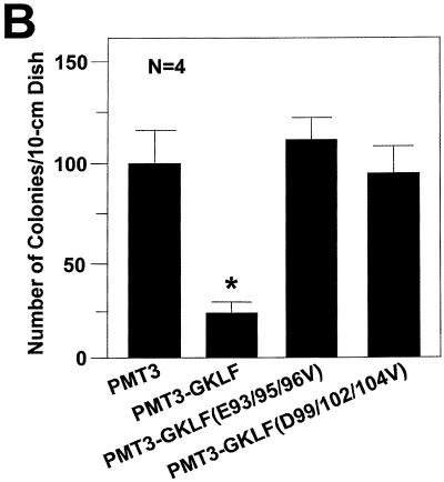
Gut-enriched Krüppel-like factor (GKLF or KLF4) is a pleiotropic (activating and repressive) transcription factor. This study characterizes the mechanisms of transactivation by GKLF. Using a GAL4 fusion assay, the activating domain of murine GKLF was localized to the 109 amino acid residues in the N-terminus. Site-directed mutagenesis showed that two adjacent clusters of acidic residues within this region are responsible for the activating effect. Transactivation by GKLF involves intermolecular interactions as demonstrated by the ability of wild-type, but not mutated, GKLF to compete with the N-terminal activation domain. In addition, wild-type adenovirus E1A, but not a mutated E1A that failed to bind p300/CBP, inhibited transactivation by the N-terminal 109 amino acids of GKLF, suggesting that p300/CBP are GKLF's interacting partners. A physical interaction between GKLF and CBP was demonstrated by glutathione- S -transferase pull-down and by in vivo co-immuno-precipitation experiments. We also showed that the two acidic amino acid clusters are essential for this interaction, since GKLF with mutations in these residues failed to co-immunoprecipitate with CBP. Importantly, the same mutations abrogated the ability of GKLF to suppress cell growth as determined by a colony suppression assay. These studies therefore provide plausible evidence for a structural and functional correlation between the transactivating and growth-suppressing effects of GKLF.


Figures

Figure 1
Figure 1
Localization of the transactivation domain of GKLF by the GAL4 fusion assay. Chinese hamster ovary (CHO) cells were co-transfected with 5 µg/10 cm dish each of the indicated effector construct and the pG5TKLUC reporter. (A) Mean fold-induction in luciferase activity by the respective effector over that by the control vector containing only the DNA-binding domain of GAL4 (effector V). Bars represent standard deviations. (B) Schematic presentation of the various GAL4 fusion effector constructs. The point mutants involving the two clusters of acidic residues, effectors 7 and 8, are drawn as E*E*E* and D*D*D*, respectively. (C) Amino acid sequence between residues 91 and 110 of GKLF and identifies the mutagenized residues. All luciferase activities were standardized to the internal control β-galactosidase activities. Data represent the means of four independent experiments, each performed in duplicate.
Figure 2
Figure 2
Localization of the transactivation domain of GKLF using its cognate binding site. CHO cells were co-transfected with 5 µg/10 cm dish each of the TDAx2-E1bTATALUC reporter and the respective effector constructs containing various regions or mutations of GKLF (effectors 17) or the vector PMT3 (V) alone. (A) Means of fold-induction in reporter activity over that of the vector (n = 4). (B) Schematic of the various effector constructs. The three E*s in effector 6 indicate the mutated glutamate residues and the D*s in effector 7 indicate the three mutated aspartate residues.
Figure 3
Figure 3
In vivo competition assays of transactivation by GKLF. CHO cells were transfected with 2 µg/10 cm dish of pG5TKLUC, 0.5 µg/10 cm dish of GAL4-GKLF(1–109) and increasing amounts of the various competitor PMT3 constructs in fold-excess relative to that of GAL4-GKLF(1–109) as indicated. The relative luciferase activity of cells that received no competitor DNA was chosen as 100%. n = 6 in experiments (A) and (B), and n = 4 in experiments (C)–(F).
Figure 3
Figure 3
In vivo competition assays of transactivation by GKLF. CHO cells were transfected with 2 µg/10 cm dish of pG5TKLUC, 0.5 µg/10 cm dish of GAL4-GKLF(1–109) and increasing amounts of the various competitor PMT3 constructs in fold-excess relative to that of GAL4-GKLF(1–109) as indicated. The relative luciferase activity of cells that received no competitor DNA was chosen as 100%. n = 6 in experiments (A) and (B), and n = 4 in experiments (C)–(F).
Figure 4
Figure 4
In vivo competition of GKLF’s transactivation by E1A-expressing constructs. CHO cells were transfected with 2 µg/10 cm dish of pG5TKLUC, 0.5 µg/10 cm dish of GAL4-GKLF(1–109) and increasing amounts of a competitor RSV construct containing either wild-type or mutated E1A in fold-excess relative to that of GAL4-GKLF(1–109). The relative luciferase activity of cells that received no competitor DNA was chosen as 100%. n = 3 in all experiments. CR indicates conserved region.
Figure 5
Figure 5
p300/CBP reverses the inhibitory effect of E1A on transactivation by GKLF. CHO cells were transfected with 2 µg/10 cm dish of pG5TKLUC, 0.5 µg/10 cm dish of GAL4-GKLF(1–109), 1 µg/10 cm dish of RSV-E1A except for lane 1, and increasing amounts of RSV-p300 (lanes 3–5) or CMV-CBP (lanes 6–8) at the indicated concentrations. The relative luciferase activity of lane 1 was taken as 100%. n = 4.
Figure 6
Figure 6
GST pull-down of GKLF by CBP. In vitro [35S]methionine-labeled GKLF was incubated with recombinant GST fusion proteins containing three different segments of CBP (lanes 4–6) or GST only (lane 3), followed by the addition of glutathione–Sepharose 4B beads. After thoroughly washing the beads with a detergent-containing solution, the bound proteins were eluted with reduced glutathione, resolved by denaturing PAGE and visualized by autoradiography. Lane 1, input GKLF, which represents 10% of the protein used in the pull-down experiments; lane 2, no input GST proteins.
Figure 7
Figure 7
Co-immunoprecipitation of GKLF and CBP. COS-1 cells were transfected with 10 µg/10 cm dish each of RSV-HA-CBP and PMT3 (lanes 1–3), wild-type (WT) PMT3-GKLF (lanes 4–6), PMT3-GKLF(E93/95/96V) (E*E*E*; lanes 7–9), or PMT3-GKLF(D99/102/104V) (D*D*D*; lanes 10–12). Twenty-four hours later, proteins were metabolically labeled with [35S]methionine in vivo. Cells were then lysed and the lysates incubated with pre-immune (PI) serum, anti-GKLF serum (αGKLF) or anti-HA antibodies (αHA). The immune complex was precipitated with protein A–Sepharose beads and resolved by gel electrophoresis following repeated washings of the beads. The asterisks identify HA-CBP. †, Wild-type GKLF; ‡, the mutant GKLF.
Figure 8
Figure 8
Colony suppression assays of GKLF. Rat1a cells were transfected with 10 µg/10 cm dish of the stated PMT3 constructs and 0.25 µg/10 cm dish of pBabe Puro (48). Two days following transfection, cells were fed media containing puromycin for 2 weeks. Resistant colonies of cells were visualized by staining with methylene blue (A). (B) Mean number of colonies for four dishes of cells transfected with each construct. *P < 0.005 by two-tailed Students’ t-test between PMT3- and PMT3-GKLF-transfected cells.
Figure 8
Figure 8
Colony suppression assays of GKLF. Rat1a cells were transfected with 10 µg/10 cm dish of the stated PMT3 constructs and 0.25 µg/10 cm dish of pBabe Puro (48). Two days following transfection, cells were fed media containing puromycin for 2 weeks. Resistant colonies of cells were visualized by staining with methylene blue (A). (B) Mean number of colonies for four dishes of cells transfected with each construct. *P < 0.005 by two-tailed Students’ t-test between PMT3- and PMT3-GKLF-transfected cells.

References

    1. Yang V.W. (1998) J. Nutr., 128, 2045–2051. - PubMed
    1. Johnson P.F. and McKnight,S.L. (1989) Annu. Rev. Biochem., 58, 799–839. - PubMed
    1. Mitchell P.J. and Tjian,R. (1989) Science, 245, 371–378. - PubMed
    1. Frankel A.D. and Kim,P.S. (1991) Cell, 65, 717–719. - PubMed
    1. McKnight S.L. and Yamamoto,K.R. (1992) Transcriptional Regulation. Cold Spring Harbor Laboratory, New York, NY.

Publication types