

Cerebral MR venography: normal anatomy and potential diagnostic pitfalls
- PMID: 10669228
- PMCID: PMC7976366
Cerebral MR venography: normal anatomy and potential diagnostic pitfalls
Abstract
Background and purpose: MR venography is often used to examine the intracranial venous system, particularly in the evaluation of dural sinus thrombosis. The purpose of this study was to evaluate the use of MR venography in the depiction of the normal intracranial venous anatomy and its variants, to assess its potential pitfalls in the diagnosis of dural venous sinus thrombosis, and to compare the findings with those of conventional catheter angiography.
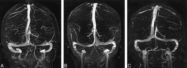
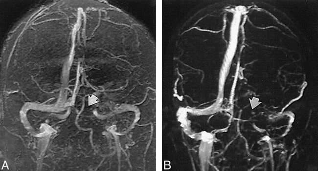
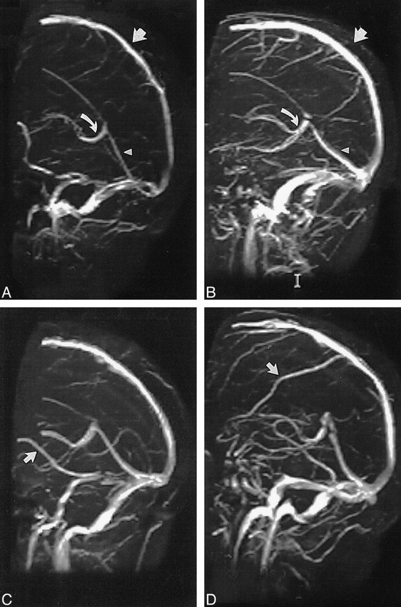
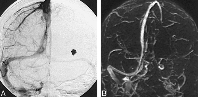
Methods: Cerebral MR venograms obtained in 100 persons with normal MR imaging studies were reviewed to determine the presence or absence of the dural sinuses and major intracranial veins.
Results: Systematic review of the 100 cases revealed transverse sinus flow gaps in 31% of the cases, with 90% of these occurring in the nondominant transverse sinus and 10% in the codominant transverse sinuses. No flow gaps occurred in the dominant transverse sinuses. The superior sagittal and straight sinuses were seen in every venogram; the occipital sinus was seen in only 10%. The vein of Galen and internal cerebral veins were also seen in every case; the basal veins of Rosenthal were present in 91%.
Conclusions: Transverse sinus flow gaps can be observed in as many as 31% of patients with normal MR imaging findings; these gaps should not be mistaken for dural sinus thrombosis.
Figures
References
-
- Yasargil MG, Damur M. Thrombosis of the cerebral veins dural sinuses. In: Newton TH, Potts DJ, eds Radiology of the Skull and Brain: Angiography St. Louis: Mosby-Year Book; 1974;2:2375-2400
-
- Casey SO, Alberico RA, Patel M, et al. Cerebral CT venography. Radiology 1996;198:163-170 - PubMed
-
- Padayachee TS, Bingham JB, Graves MJ, Colchester AC, Cox TC. Dural sinus thrombosis: diagnosis and follow-up by magnetic resonance angiography and imaging. Neuroradiology 1991;33:165-167 - PubMed
-
- Mattle HP, Wentz KU, Edelman RR, et al. Cerebral venography with MR. Radiology 1991;178:453-458 - PubMed
MeSH terms
LinkOut - more resources
Full Text Sources