

Case Reports
Apoptosis in leukoaraiosis
Affiliations
- PMID: 10669229
- PMCID: PMC7976327
Item in Clipboard
Case Reports
Apoptosis in leukoaraiosis
AJNR Am J Neuroradiol.
2000 Jan.
Abstract
We report a case of leukoaraiosis that was studied for apoptosis. In the neuropil, the number of cells that showed DNA fragmentation was 2.5 times as great in the area of leukoaraiosis as in the adjacent white matter (P = .004) and 25 times as great as in the nearby cortex (P < .001). Our findings suggest that apoptosis, predominantly of oligodendrocytes, is involved in the pathogenesis of leukoaraiosis. Within the area of leukoaraiosis, we also found numerous small veins that were partially occluded by severe collagenous thickening of the vessel walls. This collagenosis may have contributed to or resulted from chronic ischemia in that area.
Figures

Spin-echo MR image (2600/20/1 [TR/TE/excitations]) of a 1.5-cm-thick coronal slice from the frontal region of the brain. Only the left hemisphere is shown. The open arrows indicate an area of leukoaraiosis (white matter hyperintensity) that corresponds to the area that was investigated for apoptosis (see fig 2). C, cortex of the sulcus identified in fig 2
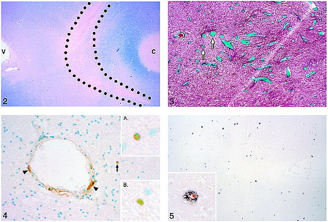
Luxol fast blue–stained section from the brain slice imaged in figure 1. The outlined area corresponds to the area of leukoaraiosis in the MR image in figure 1. Reduced Luxol fast blue staining in this area indicates demyelination. Note the darker blue staining (indicating intact myelin) of the U fibers adjacent to the cortex (C) at the bottom of a sulcus. V, lateral ventricle.fig 3. Trichrome staining shows excessively thick collagen layers (green) in the walls of small veins and venules (arrows) in the area of demyelination, where these thick-walled veins were most numerous. They are usually more numerous near the angle of the lateral ventricle.fig 4. TUNEL staining in the area of demyelination shows positive cells (brown stain) in the wall of a blood vessel (arrowheads) and in the brain parenchyma (arrow). Inset A, The brown, TUNEL-stained cell in the parenchyma appears to be apoptotic histologically in that its blue-stained nucleus is condensed and split into two segments. Inset B, Another TUNEL-stained cell in the lesional white matter with nuclear fragmentation.fig 5. A moderate number of amyloid plaques are shown in the area of cortex that was counted. Inset, Double stained for β-amyloid (black) and interleukin-1 (red) (the latter stains activated microglia and macrophages)

Comparison of the leukoaraiosis lesion (A) and an unaffected area in nearby white matter (B). Note that in the leukoaraiosis lesion, oligodendrocytes (arrows) appear to be preferentially less numerous than astrocytes, which have nuclei that are slightly larger and less densely stained (arrowheads) (hematoxylin and eosin)
Comment in
-
Staining for apoptosis: now neuropathologists can "see" leukoaraiosis.AJNR Am J Neuroradiol. 2000 Jan;21(1):42-3. AJNR Am J Neuroradiol. 2000. PMID: 10669220 Free PMC article. No abstract available.
Similar articles
-
Apoptosis in leukoaraiosis lesions.J Neurol Sci. 2002 Nov 15;203-204:169-71. doi: 10.1016/s0022-510x(02)00285-x. J Neurol Sci. 2002. PMID: 12417378
-
Cerebrovascular pathology in Alzheimer's disease and leukoaraiosis.Ann N Y Acad Sci. 2000 Apr;903:39-45. doi: 10.1111/j.1749-6632.2000.tb06348.x. Ann N Y Acad Sci. 2000. PMID: 10818487
-
Periventricular venous collagenosis: association with leukoaraiosis.Radiology. 1995 Feb;194(2):469-76. doi: 10.1148/radiology.194.2.7824728. Radiology. 1995. PMID: 7824728
-
[Age-related white matter lesions (leukoaraiosis): an update].Brain Nerve. 2013 Jul;65(7):789-99. Brain Nerve. 2013. PMID: 23832982 Review. Japanese.
-
Venous collagenosis and arteriolar tortuosity in leukoaraiosis.J Neurol Sci. 2002 Nov 15;203-204:159-63. doi: 10.1016/s0022-510x(02)00283-6. J Neurol Sci. 2002. PMID: 12417376 Review.
Cited by
-
Chronic cerebral hypoperfusion: a critical feature in unravelling the etiology of vascular cognitive impairment.Acta Neuropathol Commun. 2023 Jun 12;11(1):93. doi: 10.1186/s40478-023-01590-1. Acta Neuropathol Commun. 2023. PMID: 37309012 Free PMC article. Review.
-
Microvascular changes in the white mater in dementia.J Neurol Sci. 2009 Aug 15;283(1-2):28-31. doi: 10.1016/j.jns.2009.02.328. Epub 2009 Mar 5. J Neurol Sci. 2009. PMID: 19268311 Free PMC article.
-
Patients with vascular dementia due to microvascular pathology have significant hippocampal neuronal loss.J Neurol Neurosurg Psychiatry. 2002 Jun;72(6):747-51. doi: 10.1136/jnnp.72.6.747. J Neurol Neurosurg Psychiatry. 2002. PMID: 12023418 Free PMC article.
-
Age-related white matter changes.J Aging Res. 2011;2011:617927. doi: 10.4061/2011/617927. Epub 2011 Aug 23. J Aging Res. 2011. PMID: 21876810 Free PMC article.
-
Diffusion tensor tractography in cerebral small vessel disease: correlation with cognitive function.Neuroradiol J. 2018 Feb;31(1):83-89. doi: 10.1177/1971400916682753. Epub 2017 Oct 13. Neuroradiol J. 2018. PMID: 29027841 Free PMC article.
References
Publication types
MeSH terms
Grants and funding
LinkOut - more resources
Full Text Sources
Medical