

Proton MR spectroscopy and preoperative diagnostic accuracy: an evaluation of intracranial mass lesions characterized by stereotactic biopsy findings
- PMID: 10669230
- PMCID: PMC7976338
Proton MR spectroscopy and preoperative diagnostic accuracy: an evaluation of intracranial mass lesions characterized by stereotactic biopsy findings
Abstract
Background and purpose: MR imaging has made it easier to distinguish among the different types of intracranial mass lesions. Nevertheless, it is sometimes impossible to base a diagnosis solely on clinical and neuroradiologic findings, and, in these cases, biopsy must be performed. The purpose of this study was to evaluate the hypothesis that proton MR spectroscopy is able to improve preoperative diagnostic accuracy in cases of intracranial tumors and may therefore obviate stereotactic biopsy.
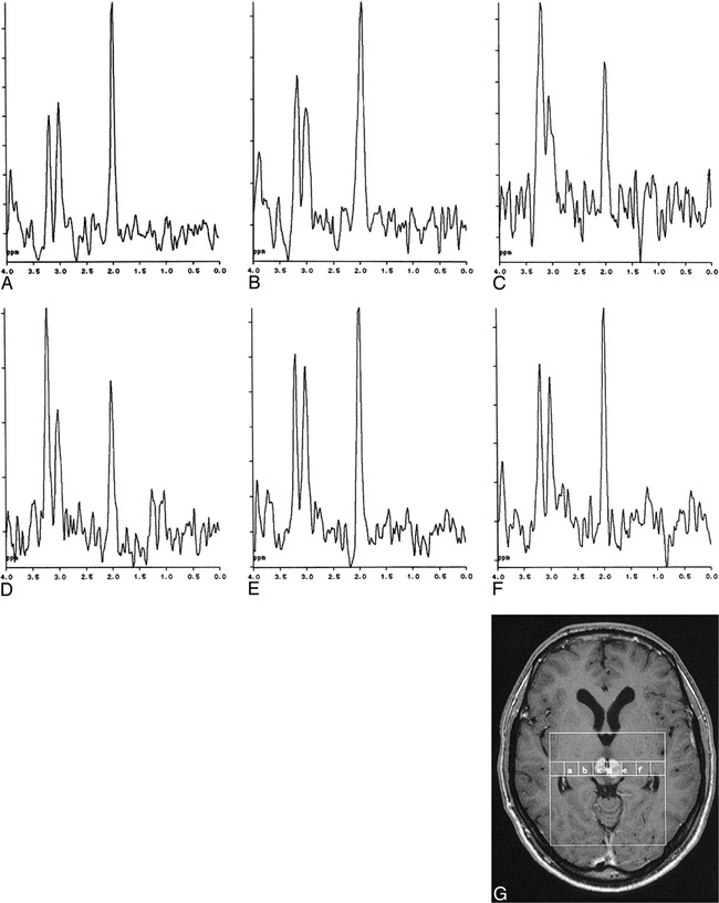
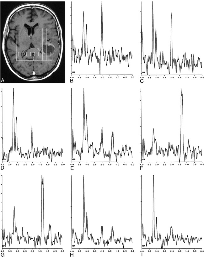
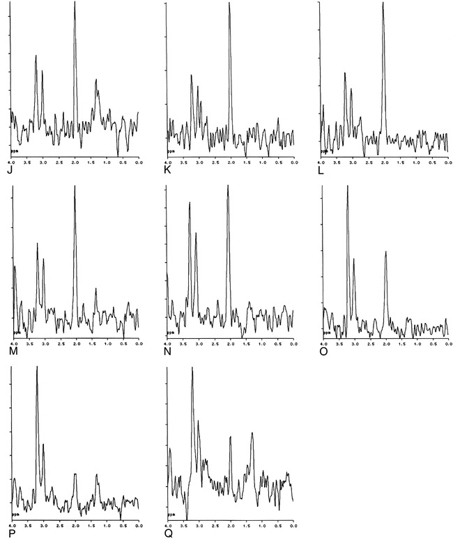
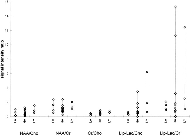
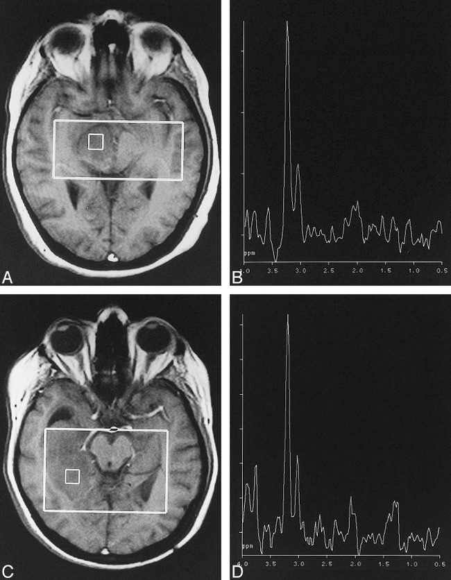
Methods: Twenty-six patients with intracranial tumors underwent MR imaging, proton MR spectroscopy, and stereotactic biopsy. MR spectroscopic findings were evaluated for the distribution pattern of pathologic spectra (NAA/Cho ratio < 1) across the lesion and neighboring tissue, for signal ratios in different tumor types, and for their potential to improve preoperative diagnostic accuracy.
Results: Gliomas and lymphomas showed pathologic spectra outside the area of contrast enhancement while four nonastrocytic circumscribed tumors (meningioma, pineocytoma, metastasis, and germinoma) showed no pathologic spectra outside the region of enhancement. No significant correlation was found between different tumor types and signal ratios. MR spectroscopy improved diagnostic accuracy by differentiating infiltrative from circumscribed tumors; however, diagnostic accuracy was not improved in terms of differentiating the types of infiltrative or circumscribed lesions.
Conclusion: MR spectroscopy can improve diagnostic accuracy by differentiating circumscribed brain lesions from histologically infiltrating processes, which may be difficult or impossible solely on the basis of clinical or neuroradiologic findings.
Figures
References
-
- Demaerel P. In vivo localized single-voxel proton magnetic resonance spectroscopy of intracranial tumors. Int J Neuroradiol 1997;3:94-110
-
- Henriksen O, Wieslander S, Gjerris F, Jensen KM. In vivo 1H-spectroscopy of human intracranial tumors at 1.5 Tesla: preliminary experience at a clinical installation. Acta Radiol 1991;32:95-99 - PubMed
-
- Demaerel P, Johannik K, Van Hecke P, et al. Localized 1H NMR spectroscopy in fifty cases of newly diagnosed intracranial tumors. J Comput Assist Tomogr 1991;15:67-76 - PubMed
-
- Sutton LN, Wehrli SL, Gennarelli L, et al. High-resolution 1H-magnetic resonance spectroscopy of pediatric posterior fossa tumors in vitro. J Neurosurg 1994;81:443-448 - PubMed
-
- Sijens PE, Knopp MV, Brunetti A, et al. 1H MR spectroscopy in patients with metastatic brain tumors: a multicenter study. Magn Reson Med 1995;33:818-826 - PubMed
Publication types
MeSH terms
LinkOut - more resources
Full Text Sources
Medical