

Visualization of intravenously administered contrast material in the CSF on fluid-attenuated inversion-recovery MR images: an in vitro and animal-model investigation
- PMID: 10669233
- PMCID: PMC7976336
Visualization of intravenously administered contrast material in the CSF on fluid-attenuated inversion-recovery MR images: an in vitro and animal-model investigation
Abstract
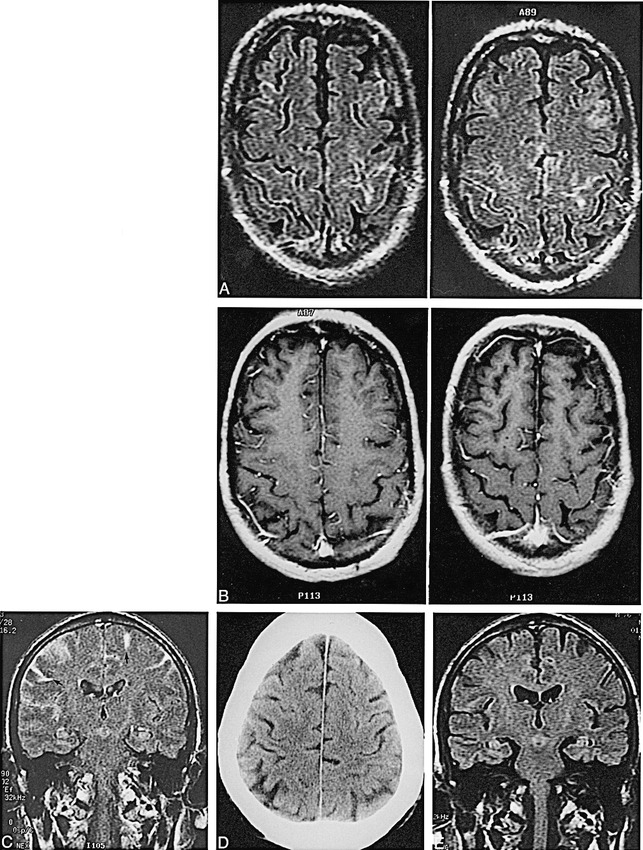
Background and purpose: The FLAIR (fluid-attenuated inversion-recovery) pulse sequence has been shown to be sensitive to abnormalities of the subarachnoid space. Our clinical experience led us to investigate whether intravenously injected contrast material can affect the appearance of the subarachnoid space on FLAIR MR images.
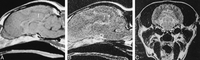
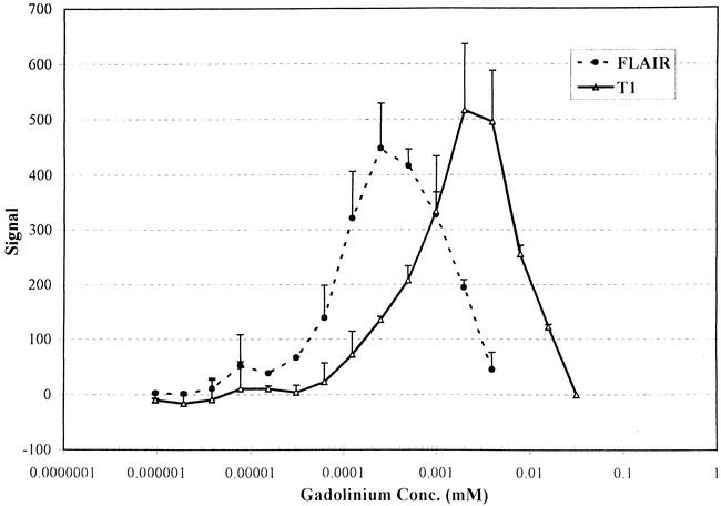
Methods: After noting unexplained high signal in the subarachnoid space on FLAIR images in a patient, we studied two dogs with sequential FLAIR MR imaging after i.v. administration of contrast material. A third dog was studied with a 6-hour delayed FLAIR sequence after triple-dose (0.3 mmol/kg) i.v. contrast administration. CSF was obtained from two animals for measurement of gadolinium concentration. A phantom was developed to determine the lowest concentration at which the effects of gadolinium were evident on FLAIR images in vitro.
Results: In all three animals, the appearance of the CSF in the ventricles or subarachnoid space was modified after administration of i.v. contrast. This was most evident on delayed images. The CSF samples showed a gadolinium concentration of 0.007 mmol/L in the dog who received the 0.1 mmol/kg dose and 0.02 mmol/L in the dog who received a triple dose. In our in vitro phantom experiments, gadolinium effects were evident on FLAIR images at a concentration four times lower than those on T1-weighted images.
Conclusion: I.v. contrast material can cross into the CSF in sufficient concentration to alter the appearance of the subarachnoid space on FLAIR images in normal dogs. Although we encountered two patients with CNS disease in whom enhancement of the CSF was seen on postcontrast FLAIR images, additional investigation is needed in humans to determine whether enhancement may occur at triple dose in healthy subjects.
Figures


References
-
- McClennan BL, Becker JA. Cerebrospinal fluid transfer of contrast material at urography. AJR Am J Roentgenol 1971;113:427-432 - PubMed
-
- Coin CG, Keranen VJ, Pennink M, Ahmad WD. Evidence of CSF enhancement in the spinal subarachnoid space after intravenous contrast medium administration: is intravenous computer assisted myelography possible? J Comput Assist Tomogr 1979;3:267-269 - PubMed
-
- Harnish PP, Northington FK, Samuel KA. Diatrizoate levels in cerebrospinal fluid following intravenous administration: role of fluid production rate. Invest Radiol 1988;23:377-380 - PubMed
-
- Knutzon RK, Poirier VC, Gerscovich EO, Brock JM, Buonocore M. The effect of intravenous gadolinium on the magnetic resonance appearance of cerebrospinal fluid. Invest Radiol 1991;26:671-673 - PubMed
Publication types
MeSH terms
Substances
LinkOut - more resources
Full Text Sources
Medical