

Phase-contrast MR imaging of the cervical CSF and spinal cord: volumetric motion analysis in patients with Chiari I malformation
- PMID: 10669242
- PMCID: PMC7976357
Phase-contrast MR imaging of the cervical CSF and spinal cord: volumetric motion analysis in patients with Chiari I malformation
Abstract
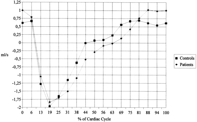
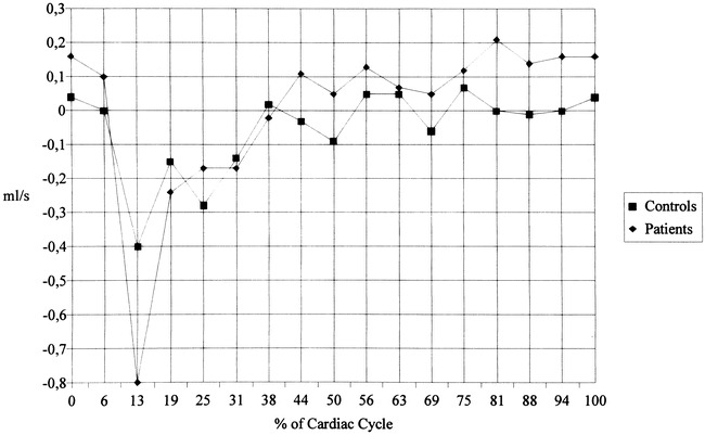
Background and purpose: Most previous MR studies of the dynamics of Chiari I malformation have been confined to sagittal images and operator-dependent measurement points in the midline. To obtain a deeper insight into the pathophysiology of the Chiari I malformation, we performed a prospective study using axial slices at the level of C2 to analyze volumetric motion data of the spinal cord and CSF over the whole cross-sectional area.
Methods: Eighteen patients with Chiari I malformation and 18 healthy control subjects underwent cardiac-gated phase-contrast imaging. Cross-sectional area measurements and volumetric flow/motion data calculations were made for the following compartments: the entire intradural space, the spinal cord, and the anterior and posterior subarachnoid space.
Results: The most striking feature was an increased early systolic caudal and diastolic cranial motion of the spinal cord in the patients. CSF pulsations in the anterior subarachnoid space were unchanged at systole but showed an impaired diastolic upward flow. In the posterior compartment, the CSF systole was slightly shortened, with an impairment of diastolic upward flow. Fourteen of the 18 patients had associated syringeal cavities. This subgroup showed an increased systolic downward displacement of the cord as compared with patients without a syrinx.
Conclusion: Obstruction of the foramen magnum in patients with Chiari I malformation causes an abrupt systolic downward displacement of the spinal cord and impairs the recoil of CSF during diastole.
Figures


Comment in
-
Toward an understanding of syringomyelia: MR imaging of CSF flow and neuraxis motion.AJNR Am J Neuroradiol. 2000 Jan;21(1):45-6. AJNR Am J Neuroradiol. 2000. PMID: 10669223 Free PMC article. No abstract available.
-
CSF flow dynamics in Chiari I malformation.AJNR Am J Neuroradiol. 2000 Sep;21(8):1564. AJNR Am J Neuroradiol. 2000. PMID: 11003298 Free PMC article. No abstract available.
References
-
- Armonda RA, Citrin CM, Foley KT, Ellenbogen RG. Quantitative cine-mode magnetic resonance imaging of Chiari I malformations: an analysis of cerebrospinal fluid dynamics. Neurosurgery 1994;35:214-224 - PubMed
-
- Bhadelia RA, Bogdan AR, Wolpert SM, Lev S, Appignani BA, Heilman CB. Cerebrospinal fluid flow waveform: analysis in patients with Chiari I malformation by means of gated phase-contrast MR imaging velocity measurements. Radiology 1995;196:195-202 - PubMed
-
- Oldfield EH, Muraszko K, Shawker TH, Patronas NJ. Pathophysiology of syringomyelia associated with Chiari I malformation of the cerebellar tonsils. J Neurosurg 1994;80:3-15 - PubMed
-
- Terae S, Miyasaka K, Abe S, Abe H, Tashiro K. Increased pulsatile movement of the hindbrain in syringomyelia associated with the Chiari malformation: cine-MR with presaturation bolus tracking. Neuroradiology 1994;36:125-129 - PubMed
MeSH terms
LinkOut - more resources
Full Text Sources
Other Literature Sources
Medical
Miscellaneous