

Masticator space abnormalities associated with mandibular osteoradionecrosis: MR and CT findings in five patients
- PMID: 10669246
- PMCID: PMC7976370
Masticator space abnormalities associated with mandibular osteoradionecrosis: MR and CT findings in five patients
Abstract
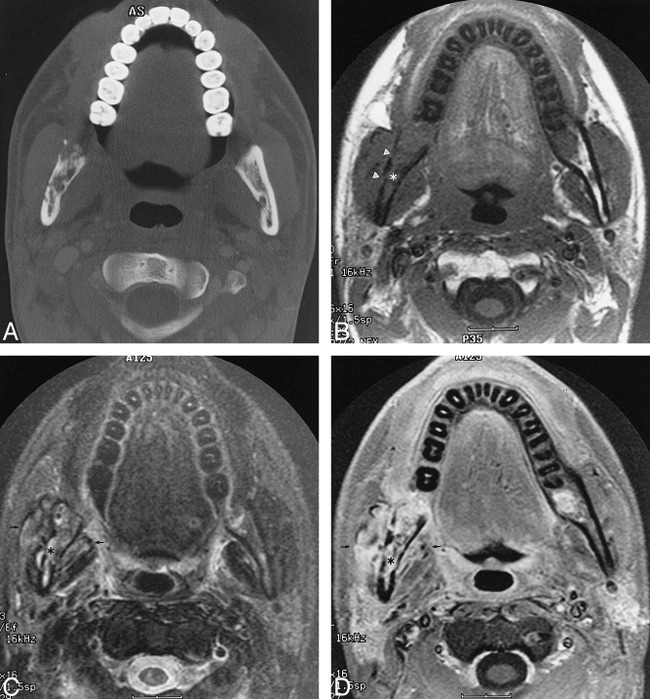
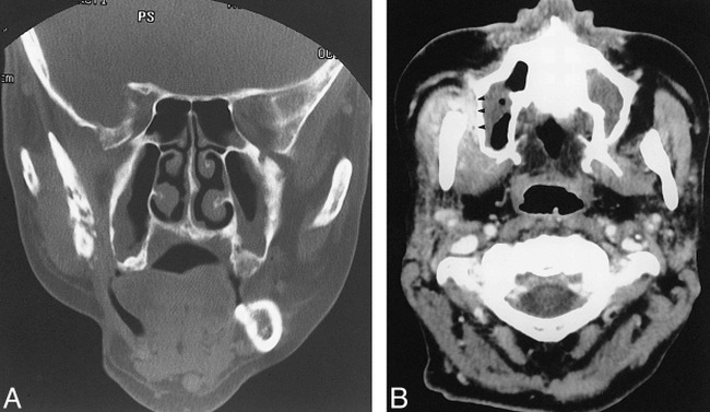
Background and purpose: Imaging of patients with a clinical diagnosis of mandibular osteoradionecrosis (ORN) is often performed to support that clinical suspicion, evaluate the extent of the disease, or exclude coexistent tumor recurrence. The purpose of our study was to describe the clinical, MR imaging, and CT features of five patients with mandibular ORN associated with prominent soft-tissue abnormality in the adjacent masticator muscles.
Methods: The MR and CT examinations of five patients with mandibular ORN associated with soft-tissue abnormalities in the adjacent masticator muscles were reviewed. All patients had received external beam radiotherapy for primary head and neck malignancies, with a total radiation dose range of 60 Gy to 69 Gy in 30 to 38 fractions.
Results: CT revealed the typical osseous findings of cortical disruption, trabecular disorganization, and fragmentation in all five patients. Abnormal diffuse enhancement of the adjacent masseter and pterygoid muscles was noted in all patients. Four patients had prominent mass-like thickening of these muscles adjacent to the osseous abnormality. Of the three patients who underwent MR imaging, all showed homogeneous abnormal T1 hypointensity, T2 hyperintensity, and intense enhancement of the bone marrow in the involved mandible. The masticator muscles adjacent to the osseous abnormality also showed abnormal T2 hyperintensity and intense diffuse enhancement on MR images.
Conclusion: Mandibular ORN can be associated with prominent soft-tissue thickening and enhancement in the adjacent musculature. These changes can appear mass-like and are not related to tumor recurrence or metastatic disease.
Figures
Similar articles
-
The normal and diseased masticator space.Semin Ultrasound CT MR. 1990 Dec;11(6):476-85. Semin Ultrasound CT MR. 1990. PMID: 2275809 No abstract available.
-
Dynamic MR imaging of mandibular osteoradionecrosis.Acta Radiol. 2000 Jan;41(1):31-7. Acta Radiol. 2000. PMID: 10665867
-
MRI findings in patients with severe trismus following radiotherapy for nasopharyngeal carcinoma.Eur Radiol. 2009 Nov;19(11):2586-93. doi: 10.1007/s00330-009-1445-z. Epub 2009 Jun 6. Eur Radiol. 2009. PMID: 19504110
-
Osteoradionecrosis of the mandible: through a radiologist's eyes.Clin Radiol. 2015 Feb;70(2):197-205. doi: 10.1016/j.crad.2014.09.012. Epub 2014 Oct 18. Clin Radiol. 2015. PMID: 25446325 Review.
-
Masticator space: CT and MRI of secondary tumor spread.AJR Am J Roentgenol. 2007 Aug;189(2):488-97. doi: 10.2214/AJR.07.2212. AJR Am J Roentgenol. 2007. PMID: 17646477 Review.
Cited by
-
Sclerotic Lesions of the Jaw: A Pictorial Review.J Belg Soc Radiol. 2021 Apr 8;105(1):21. doi: 10.5334/jbsr.2208. J Belg Soc Radiol. 2021. PMID: 33870085 Free PMC article.
-
Radiolucent lesions of the mandible: a pattern-based approach to diagnosis.Insights Imaging. 2014 Feb;5(1):85-101. doi: 10.1007/s13244-013-0298-9. Epub 2013 Dec 10. Insights Imaging. 2014. PMID: 24323536 Free PMC article.
-
Imaging of the Posttreatment Head and Neck: Expected Findings and Potential Complications.Radiol Imaging Cancer. 2024 Jan;6(1):e230155. doi: 10.1148/rycan.230155. Radiol Imaging Cancer. 2024. PMID: 38276904 Free PMC article. Review.
-
Identification and management of recurrent oral squamous cell carcinoma in the clinical presentation of osteoradionecrosis: a single-center case series for treatment experience sharing.BMC Oral Health. 2025 Feb 13;25(1):228. doi: 10.1186/s12903-025-05603-4. BMC Oral Health. 2025. PMID: 39948512 Free PMC article.
-
Imaging of Surgical Free Flaps in Head and Neck Reconstruction.AJNR Am J Neuroradiol. 2019 Jan;40(1):5-13. doi: 10.3174/ajnr.A5776. Epub 2018 Nov 8. AJNR Am J Neuroradiol. 2019. PMID: 30409846 Free PMC article. Review.
References
-
- MacDougall JA, Evans AM, Lindsay RK. Osteoradionecrosis of the mandible and its treatment. Am J Surg 1963;106:816-818 - PubMed
-
- Bedwinek JM, Shukovsky LJ, Fletcher GH, Daley TE. Osteonecrosis in patients treated with definitive radiotherapy for squamous cell carcinomas of the oral cavity and naso- and oropharynx. Radiology 1976;119:665-667 - PubMed
-
- Morrish RB, Chan E, Silverman S, Meyer J, Fu KK, Greenspan D. Osteonecrosis in patients irradiated for head and neck carcinoma. Cancer 1981;47:1980-1983 - PubMed
-
- Murray CG, Herson J, Daly TE, Zimmerman S. Radiation necrosis of the mandible: a 10-year study: part I: factors influencing the onset of necrosis. Int J Radiat Oncol Biol Phys 1980;6:543-548 - PubMed
-
- Epstein JB, Wong FLW, Stevenson-Moore P. Osteoradionecrosis: clinical experience and a proposal for classification. J Oral Maxillofac Surg 1987;45:104-110 - PubMed
MeSH terms
LinkOut - more resources
Full Text Sources
Medical