. 2000 Mar;20(5):1526-36.
doi: 10.1128/MCB.20.5.1526-1536.2000.

Molecular mechanism for the Shp-2 tyrosine phosphatase function in promoting growth factor stimulation of Erk activity

Affiliations

Molecular mechanism for the Shp-2 tyrosine phosphatase function in promoting growth factor stimulation of Erk activity

Z Q Shi et al. Mol Cell Biol. 2000 Mar.

Abstract

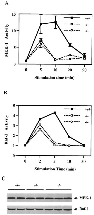
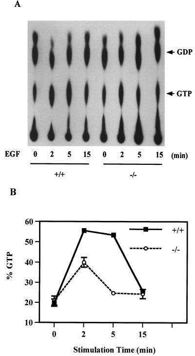
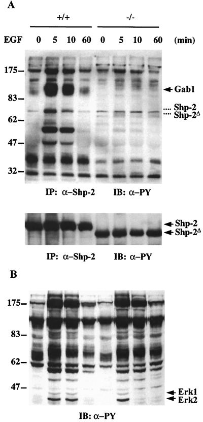
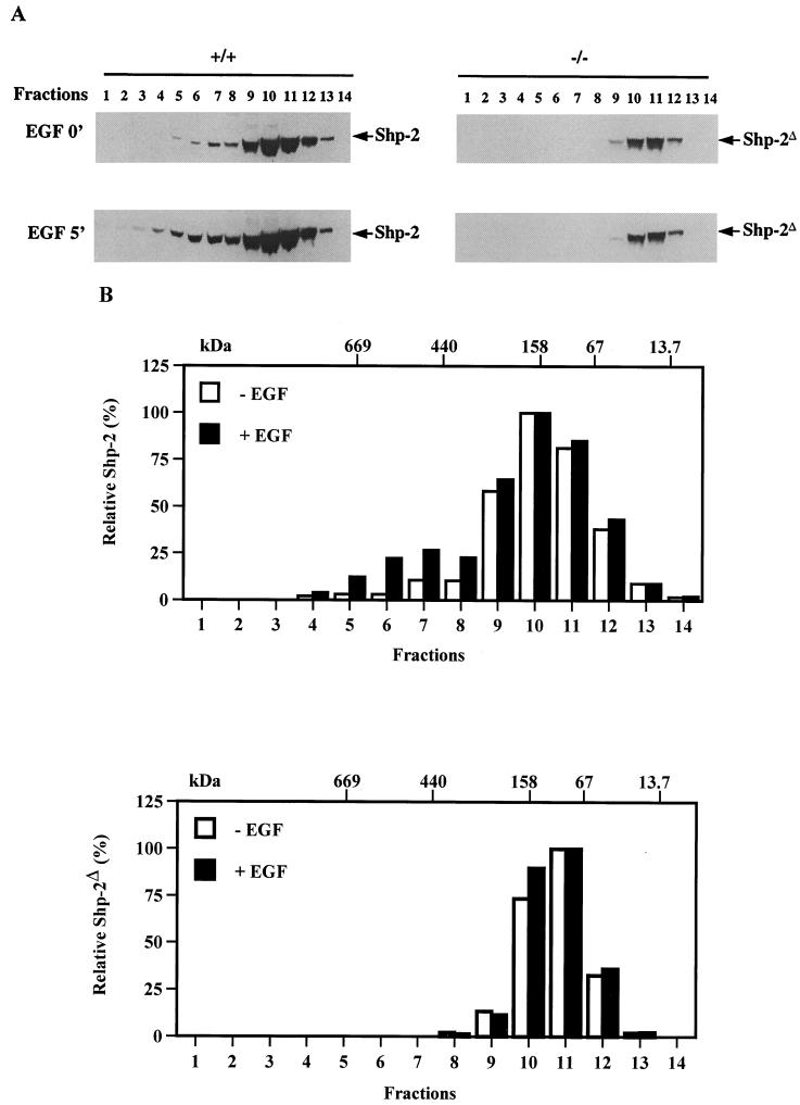
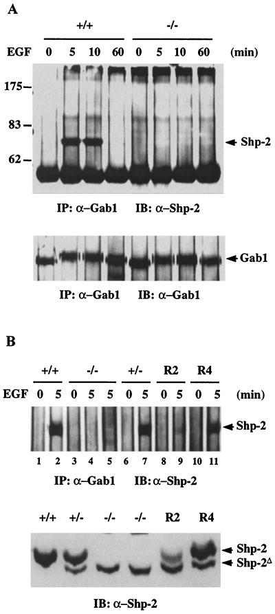
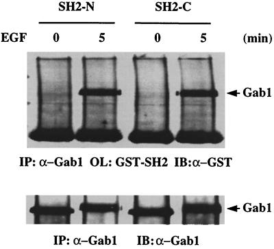
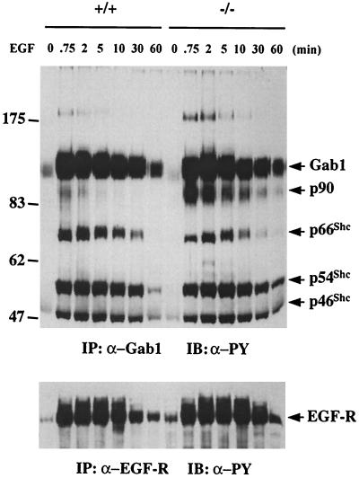
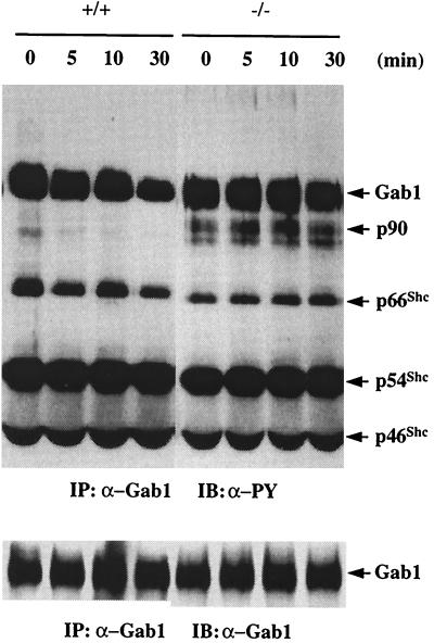
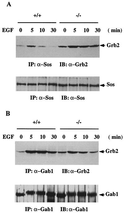
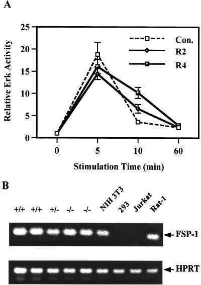
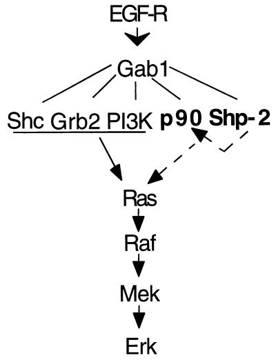
We have previously shown that activation of extracellular signal-regulated kinase (Erk) by epidermal growth factor (EGF) treatment was significantly decreased in mouse fibroblast cells expressing a mutant Shp-2 molecule lacking 65 amino acids in the SH2-N domain, Shp-2(Delta46-110). To address the molecular mechanism for the positive role of Shp-2 in mediating Erk induction, we evaluated the activation of signaling components upstream of Erk in Shp-2 mutant cells. EGF-stimulated Ras, Raf, and Mek activation was significantly attenuated in Shp-2 mutant cells, suggesting that Shp-2 acts to promote Ras activation or to suppress the down-regulation of activated Ras. Biochemical analyses indicate that upon EGF stimulation, Shp-2 is recruited into a multiprotein complex assembled on the Gab1 docking molecule and that Shp-2 seems to exert its biological function by specifically dephosphorylating an unidentified molecule of 90 kDa in the complex. The mutant Shp-2(Delta46-110) molecule failed to participate in the Gab1-organized complex for dephosphorylation of p90, correlating with a defective activation of the Ras-Raf-Mek-Erk cascade in EGF-treated Shp-2 mutant cells. Evidence is also presented that Shp-2 does not appear to modulate the signal relay from EGF receptor to Ras through the Shc, Grb2, and Sos proteins. These results begin to elucidate the mechanism of Shp-2 function downstream of a receptor tyrosine kinase to promote the activation of the Ras-Erk pathway, with potential therapeutic applications in cancer treatment.


Figures

FIG. 1
FIG. 1
Reduced activation of Mek and Raf in Shp-2 mutant cells. Fibroblasts of one wild-type (+/+) and two homozygous Shp-2 mutant (−/−) cell lines were starved in serum-free DMEM medium for 36 h and then stimulated with EGF (100 ng/ml) for indicated times. Mek (A) and Raf (B) kinase activities were measured by a coupled in vitro kinase assay as described in the text. Incorporation of 32P into MBP was determined by scintillation counting to reflect the kinase activities. The protein expression levels of Mek-1 and Raf-1 in two wild-type (+/+), two heterozygous (+/−), and three homozygous mutant (−/−) cell lines were evaluated by immunoblot analysis using specific antibodies against Mek-1 and Raf-1, respectively (C).
FIG. 2
FIG. 2
Decreased Ras activation in Shp-2 mutant cells. Ras protein was immunoprecipitated from 32P-labeled control and EGF-stimulated cell lysates. Following extensive washes with cell lysis buffer, GTP and GDP were eluted from the precipitates and resolved by TLC followed by visualization with autoradiography on X-ray film (A). GTP and GDP spots were removed from the TLC plate and counted in a Beckman scintillation counter. The ratio GTP/(GDP + GTP) was calculated to represent Ras activity (B). Data were averaged from samples of three independent experiments.
FIG. 3
FIG. 3
EGF-induced association of Shp-2 with tyrosine-phosphorylated proteins. (A) Shp-2 protein was immunoprecipitated (IP) from wild-type (+/+) or mutant (−/−) cell lysates, resolved by SDS-PAGE, and immunoblotted (IB) with anti-PY antibody. The same membrane was then stripped and reprobed with an anti-Shp-2 antibody raised against the C-terminal region of Shp-2. The corresponding positions of wild-type and mutant Shp-2 bands were indicated with dashed lines in the upper panel. (B) Equal amounts of whole cell lysates from control or EGF-stimulated cells were resolved by SDS-PAGE, transferred to a nitrocellulose membrane, and blotted with an anti-PY antibody.
FIG. 4
FIG. 4
Distinct protein complexes of wild-type and mutant Shp-2 in response to EGF stimulation. Control and EGF-treated whole cell lysates were applied on a Superose 6 HR10/30 gel filtration column. Trichloroacetic acid-precipitated proteins from each fraction were separated by SDS-PAGE and immunoblotted with an anti-Shp-2 antibody. Estimated molecular masses were calculated based on the standard curve produced from the molecular weight markers described in the text. (A) Immunoblotting results; (B) densitometry scanning results of the immunoblots shown in panel A.
FIG. 5
FIG. 5
Coimmunoprecipitation of Gab1 with wild-type but not mutant Shp-2. (A) Anti-Gab1 antiserum (2 μl) was added to 1 ml (1 mg of total protein) of cell lysates from Shp-2+/+ or Shp-2−/− cells stimulated by EGF at the indicated times and immunoprecipitated (IP). The precipitates were immunoblotted (IB) with an anti-Shp-2 antibody that recognizes both wild-type and Shp-2Δ46-110 proteins. The same membrane was then reprobed with an anti-Gab1 antiserum. (B) Gab1 protein was immunoprecipitated from EGF-stimulated wild-type (+/+), heterozygous (+/−), homozygous mutant (−/−), and two rescue cell lines, R2 and R4, in which the wild-type Shp-2 cDNA was reintroduced (55). Each immunoprecipitation was performed with 800 μg of total protein except in lane 5, which had 3 mg of total protein. In the lower panel, equal amounts (40 μg) of cell lysates were directly immunoblotted with the anti-Shp-2 antibody.
FIG. 6
FIG. 6
Direct recognition of tyrosine-phosphorylated Gab1 by Shp-2 SH2 domains. Gab1 protein was immunoprecipitated (IP) from control or EGF-stimulated wild-type cell lysates. The immunocomplex was resolved by SDS-PAGE, transferred to a nitrocellulose membrane, and overlaid (OL) with the SH2-N or SH2-C GST fusion protein. Binding of the GST fusion proteins was detected by immunoblotting (IB) the membrane with anti-GST antibody.
FIG. 7
FIG. 7
Functional interaction between Gab1 and Shp-2. Total cell lysates (1 mg of total proteins) from Shp-2+/+ or Shp-2−/− cells stimulated by 100 ng of EGF per ml for the indicated times were mixed with 2 μl of anti-Gab1 antibody and protein A-Sepharose 4B beads for immunoprecipitation (IP). The precipitates were then immunoblotted (IB) with anti-PY antibody to assess the tyrosine phosphorylation levels of Gab1 as well as Gab1-associated phosphoproteins. In the lower panel, the EGF-R was immunoprecipitated from the same cell lysates and immunoblotted with anti-PY antibody.
FIG. 8
FIG. 8
Dephosphorylation of Gab1 and Gab1-associated proteins by Shp-2 in vitro. Shp-2+/+ and Shp-2−/− cells were stimulated by 100 ng of EGF per ml for 45 s and lysed in cell lysis buffer; Gab1 was immunoprecipitated (IP) from the cell lysates. The precipitates were washed twice with HNTG buffer without phosphatase inhibitors and, twice with phosphatase buffer (50 mM imidazole [pH 7.5], 10 mM DTT, 5 mM EDTA) and then incubated in 20 μl of phosphatase buffer for 5, 10, and 30 min at 30°C. The reaction was stopped by 5 μl of 5× SDS sample buffer, immunoblotted (IB) by anti-PY antibody, and then reprobed with anti-Gab1 antibody.
FIG. 9
FIG. 9
Alternative binding of Grb2-Gab1 and Grb2-Sos. (A) Sos-1 was immunoprecipitated (IP) from control or EGF-stimulated cell lysates (1 mg of total proteins) and immunoblotted (IB) with an anti-Grb2 antibody. The same membrane was stripped and reprobed by the anti-Sos antibody to determine the amount of Sos protein precipitated from each sample. (B) Immunoprecipitation was performed with an anti-Gab1 antibody, blotted with an anti-Grb2 antibody, and then reprobed with anti-Gab1.
FIG. 10
FIG. 10
Rescue of defective Erk induction by EGF in Shp-2−/− cells and RT-PCR analysis of the fsp-1 mRNA expression. (A) Vector-transfected control cell line (Con.) and two cell clones expressing different levels of wild-type Shp-2 were stimulated with EGF (100 ng/ml) for indicated times. Erk protein was immunoprecipitated from cell lysates, and the kinase activity was measured by the in vitro kinase assay as described previously (45). (B) Total RNA of different cell lines was purified from 3 × 10⁶ cells by using an RNeasy Mini kit from Qiagen as specified by the manufacturer. Gene-specific primers for mouse fsp-1 (sense primer, 5′-CAG CGA AAG AGG GTG ACA AGT TCA-3′; antisense primer, 5′-ATG TGC GAA GAA GCC AGA GTA AGG-3′) (46, 49) or hypoxanthine phosphoribosyltransferase (HPRT), as a positive control (38), were used to amplify 1 μg of RNA, using a GeneAmp RNA PCR kit (Perkin-Elmer) as instructed by the manufacturer. RT-PCR products were resolved in 1.2% agarose gel and visualized by ethidium bromide staining.
FIG. 11
FIG. 11
A model of Shp-2 function. It has been known that Shc, Grb2, and phosphoinositol 3-kinase (PI3K) are associated with Gab1 and are involved in mediating Ras activation following EGF treatment. Results from this work allow us to propose a novel mechanism for the role of Shp-2 in promoting the activation of the Ras-Erk pathway by EGF, possibly through dephosphorylation of Gab1-associated p90, which is indicated by arrows with dashed lines.

References

    1. Allard J D, Chang H C, Herbst R, McNeill H, Simon M A. The SH2-containing tyrosine phosphatase corkscrew is required during signaling by sevenless, Ras1 and Raf. Development. 1996;122:1137–1146. - PubMed
    1. Barnard D, Diaz B, Clawson D, Marshall M. Oncogenes, growth factors and phorbol esters regulate Raf-1 through common mechanisms. Oncogene. 1998;17:1539–1547. - PubMed
    1. Bennett A M, Hausdorff S F, O'Reilly A M, Freeman R M, Neel B G. Multiple requirements for SHPTP2 in epidermal growth factor-mediated cell cycle progression. Mol Cell Biol. 1996;16:1189–1202. - PMC - PubMed
    1. Bennett A M, Tang T L, Sugimoto S, Walsh C T, Neel B G. Protein-tyrosine-phosphatase SHPTP2 couples platelet-derived growth factor receptor beta to Ras. Proc Natl Acad Sci USA. 1994;91:7335–7339. - PMC - PubMed
    1. Buday L, Downward J. Epidermal growth factor regulates p21ras through the formation of a complex of receptor, Grb2 adapter protein, and Sos nucleotide exchange factor. Cell. 1993;73:611–620. - PubMed

Publication types

MeSH terms

Substances