. 2000 Mar;20(5):1537-45.
doi: 10.1128/MCB.20.5.1537-1545.2000.

Nck-interacting Ste20 kinase couples Eph receptors to c-Jun N-terminal kinase and integrin activation

Affiliations

Nck-interacting Ste20 kinase couples Eph receptors to c-Jun N-terminal kinase and integrin activation

E Becker et al. Mol Cell Biol. 2000 Mar.

Abstract

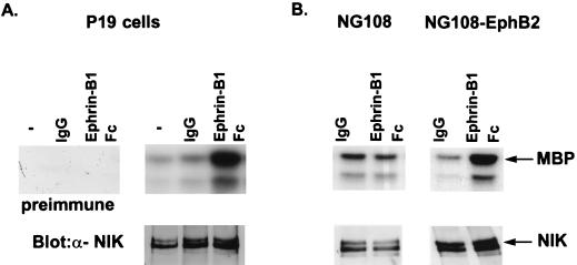
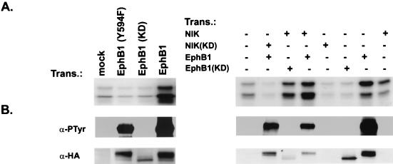
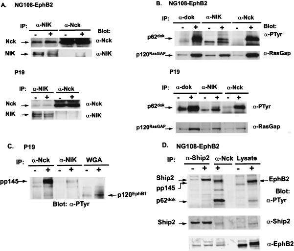
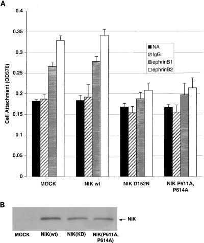
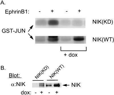
The mammalian Ste20 kinase Nck-interacting kinase (NIK) specifically activates the c-Jun amino-terminal kinase (JNK) mitogen-activated protein kinase module. NIK also binds the SH3 domains of the SH2/SH3 adapter protein Nck. To determine whether Nck functions as an adapter to couple NIK to a receptor tyrosine kinase signaling pathway, we determined whether NIK is activated by Eph receptors (EphR). EphRs constitute the largest family of receptor tyrosine kinases (RTK), and members of this family play important roles in patterning of the nervous and vascular systems. In this report, we show that NIK kinase activity is specifically increased in cells stimulated by two EphRs, EphB1 and EphB2. EphB1 kinase activity and phosphorylation of a juxtamembrane tyrosine (Y594), conserved in all Eph receptors, are both critical for NIK activation by EphB1. Although pY594 in the EphB1R has previously been shown to bind the SH2 domain of Nck, we found that stimulation of EphB1 and EphB2 led predominantly to a complex between NIK/Nck, p62(dok), RasGAP, and an unidentified 145-kDa tyrosine-phosphorylated protein. Tyrosine-phosphorylated p62(dok) most probably binds directly to the SH2 domain of Nck and RasGAP and indirectly to NIK bound to the SH3 domain of Nck. We found that NIK activation is also critical for coupling EphB1R to biological responses that include the activation of integrins and JNK by EphB1. Taken together, these findings support a model in which the recruitment of the Ste20 kinase NIK to phosphotyrosine-containing proteins by Nck is an important proximal step in the signaling cascade downstream of EphRs.


Figures

FIG. 1
FIG. 1
Immunoblot and immune complex kinase assay of NIK from P19 and NG108-EphB2 cells stimulated with ephrinB1. Lysates from P19 cells (A) or NG108-EphB2 cells (B) that were unstimulated (−), treated with clustering antibody alone (IgG), or stimulated with preclustered ephrin (Ephrin-B1 Fc) were immunoprecipitated with anti-NIK antibodies. Half the immunoprecipitates were subjected to an in vitro kinase assay using [γ-32P]ATP and myelin basic protein (MBP) as the substrate. Reaction products were separated by SDS-PAGE (12.5% polyacrylamide) and visualized by autoradiography (upper panels). Half of the immunoprecipitate was immunoblotted with the anti-NIK antibodies, to ensure equal levels of NIK expression (lower panels). The anti-NIK antibody (α-NIK) recognizes two isoforms of NIK that run at apparent molecular masses of 140 and 150 kDa, possibly due to alternate splicing of NIK. To control for nonspecific kinase activity coimmunoprecipitating with the anti-NIK antibodies, an immune complex kinase assay was performed on P19 cell lysates using preimmune serum (A). In addition, to demonstrate that the increase in NIK activity in stimulated NG108-EphB2 cells required the EphB2 receptor, NIK activity was assessed in the parental NG108 cell line (B).
FIG. 2
FIG. 2
NIK activity in 293 cells transfected (Trans.) with wild-type and mutant EphB1 receptors. (A) 293 cells were transfected with expression plasmids containing different cDNAs corresponding to the proteins as indicated. To assess NIK activation, total-protein extracts were immunoprecipitated with anti-NIK antibodies and subjected to an in vitro kinase assay as described in the legend to Fig. 1. (B) Transfected lysates were immunoblotted with antibodies to the HA epitope (12CA5) (α-HA) to control for EphB1 expression (EphB1 was HA epitope tagged). To assess expression of EphB1, the same extracts were precipitated with wheat germ agglutinin and tyrosine-phosphorylated EphB1 was determined by immunoblotting the washed precipitates with antiphosphotyrosine antibodies (α-PTyr).
FIG. 3
FIG. 3
Proteins associated with NIK in unstimulated or ephrinB1-stimulated P19 and NG108-EphB2 cells. Cells were treated for 15 to 20 min either with preclustered ephrinB1 (+) or with clustering antibody alone (−). Lysates from P19 or NG108-EphB2 cells were immunoprecipitated (IP) with anti-Nck (α-Nck), anti-NIK (α-NIK), anti-p62dok (α-dok), or anti-SHIP2 antibodies as indicated. (For immunoprecipitation, NIK antibodies were covalently coupled to protein A-Sepharose beads). The washed immunoprecipitates were separated by SDS-PAGE (10% polyacrylamide or as otherwise specified) and, after being transferred to nitrocellulose filters, probed with antibodies as indicated. In panel C, EphB1 was precipitated using wheat germ agglutinin (WGA). In panel D, after the nitrocellulose filter was probed with antiphosphotyrosine (α-PTyr) antibodies, the filter was stripped and reprobed first with anti-SHIP2 antibodies and then with anti-EphB2 antibodies. (A) Antibodies to NIK (α-NIK) coimmunoprecipitate Nck in both unstimulated and ephrinB1-stimulated cells. (B) In stimulated cells, endogenous NIK and Nck coimmunoprecipitate with tyrosine-phosphorylated p62dok and RasGAP which is not tyrosine phosphorylated (data not shown). (C and D) Antibodies to NIK (α-NIK) and Nck (α-Nck) immunoprecipitate an unidentified tyrosine-phosphorylated protein of 145 kDa (pp145), which is not the EphB1 or EphB2 receptor. In panel C, the tyrosine-phosphorylated EphB1 receptor was detected by immunoblotting WGA precipitates with antiphosphotyrosine antibodies as described in the legend to Fig. 2B. The tyrosine-phosphorylated protein at 145 kDa runs at a higher molecular mass than the EphB1 receptor. In stimulated EphB2 cells, pp145, which coimmunoprecipitates with anti-Nck antibodies, is not the EphB2 receptor, since pp145 does not immunoblot with antibodies to EphB2 (D). In panel D, the filter was first probed with antiphosphotyrosine antibodies and, after being stripped, was reprobed with anti-SHIP2 (α-Ship2) and then anti-EphB2 antibodies. The residual bands present in the anti-SHIP2 and the anti-EphB2 blot reflect incomplete stripping of the previously used antibodies. Differences in the concentration of the polyacrylamide gel, 7% (C) versus 10% (D), account for the apparent differences in migration between pp145 and EphB1 compared with pp145 and EphB2. (D) Antibodies to SHIP2 immunoprecipitate tyrosine-phosphorylated SHIP2 from NG108-EphB2 cells stimulated with ephrinB1. pp145 is not SHIP2, since we cannot immunoblot pp145 with antibodies to SHIP2.
FIG. 4
FIG. 4
Solid-phase attachment assay of P19 cells to fibrinogen. (A) P19 cells were transiently transfected with 6 μg of NIK(WT) or mutant NIK(KD), as shown on the x axis. At 48 h after transfection, solid-phase attachment assays (see Materials and Methods) were conducted on fibrinogen-coated plates displaying ephrinB1 or ephrinB2, clustering antibody alone (IgG), or no addition (NA). Adherent cells were stained with crystal violet and quantified by measurement of the optical density at 570 nm (OD570). ∗, P < 0.001; ∗∗, P < 0.01. (B) To confirm the presence of equal levels of NIK expression, lysates were immunoprecipitated with the anti-Myc antibody 9E10 and immunoblotted with the same antibody.
FIG. 5
FIG. 5
EphrinB1-stimulated JNK activation in P19 cells that inducibly express NIK(KD). (A) Stable P19 cell lines that inducibly express HA-tagged NIK(KD) or NIK(WT) were isolated as described in Materials and Methods. To determine whether expression of NIK(KD) or NIK(WT) affects JNK activation by ephrinB1, P19 cell lines growing with (+ dox) or without doxycycline were treated with preclustered ephrin B1 (+) or the clustering IgG alone (−) for 20 min. To assay JNK activity, lysates were incubated with 10 μg of GST-Jun, and after they were washed, an in vitro kinase assay was performed as described previously (43). Reaction products were separated by SDS-PAGE (15% polyacrylamide) and visualized by autoradiography. (B) NIK(KD) and NIK(WT) expression induced by doxycycline was measured by immunoblotting 50 μg of lysates from P19 cells treated for 48 h in the absence (−) or presence (+) of 1 μg of doxycycline (dox) per ml with anti-NIK antibodies (α:NIK).

References

    1. Adams R H, Wilkinson G A, Weiss C, Diella F, Gale N W, Deutsch U, Risau W, Klein R. Roles of ephrinB ligands and EphB receptors in cardiovascular development: demarcation of arterial/venous domains, vascular morphogenesis, and sprouting angiogenesis. Genes Dev. 1999;13:295–306. - PMC - PubMed
    1. Bohme B, VandenBos T, Cerretti D P, Park L S, Holtrich U, Rubsamen-Waigmann H, Strebhardt K. Cell-cell adhesion mediated by binding of membrane-anchored ligandLERK-2 to the EPH-related receptor human embryonal kinase 2 promotes tyrosine kinase activity. J Biol Chem. 1996;217:24747–24752. - PubMed
    1. Braverman L E, Quilliam L A. Identification of Grb4/Nckbeta, a src homology 2 and 3 domain-containing adapter protein having similar binding and biological properties to Nck. J Biol Chem. 1999;274:5542–5549. - PubMed
    1. Bruckner K, Pasquale E B, Klein R. Tyrosine phosphorylation of transmembrane ligands for Eph receptors. Science. 1997;275:1640–1643. - PubMed
    1. Carpino N, Wisniewski D, Strife A, Marshak D, Kobayashi R, Stillman B, Clarkson B. p62(dok): a constitutively tyrosine-phosphorylated, GAP-associated protein in chronic myelogenous leukemia progenitor cells. Cell. 1997;88:197–204. - PubMed

Publication types

MeSH terms

Substances