. 2000 Mar;20(5):1649-58.
doi: 10.1128/MCB.20.5.1649-1658.2000.

CIZ, a zinc finger protein that interacts with p130(cas) and activates the expression of matrix metalloproteinases

Affiliations

CIZ, a zinc finger protein that interacts with p130(cas) and activates the expression of matrix metalloproteinases

T Nakamoto et al. Mol Cell Biol. 2000 Mar.

Abstract

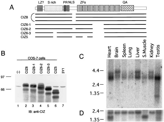
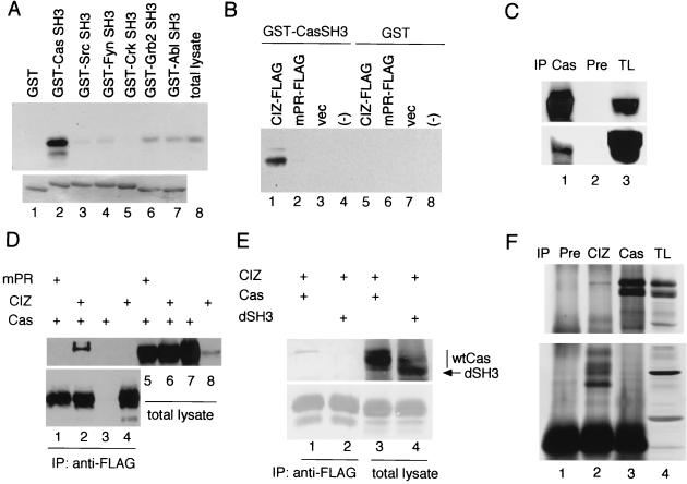
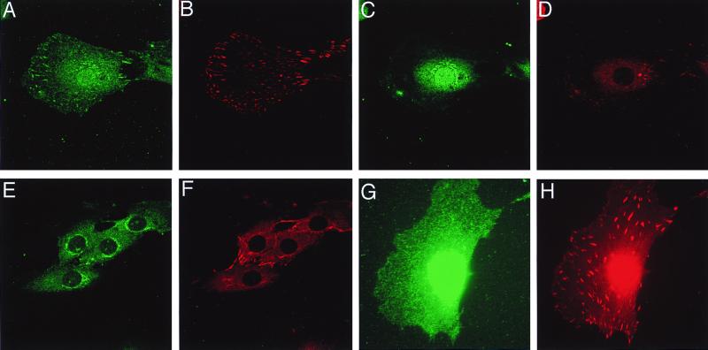
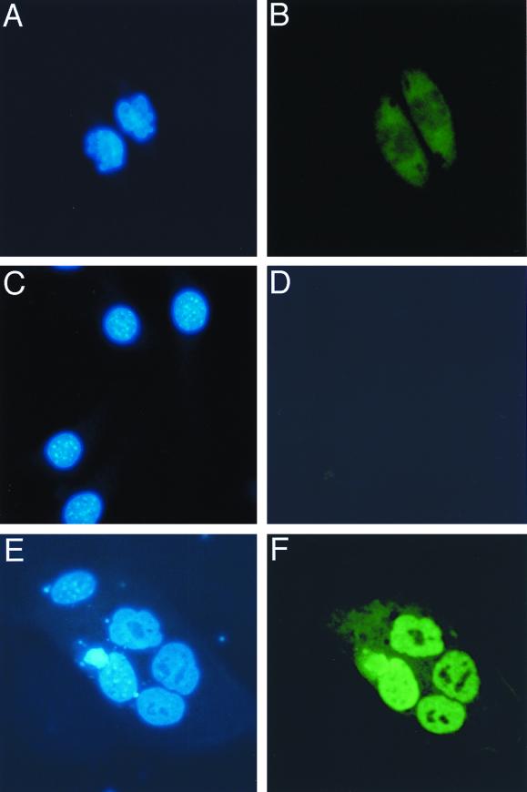
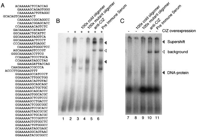
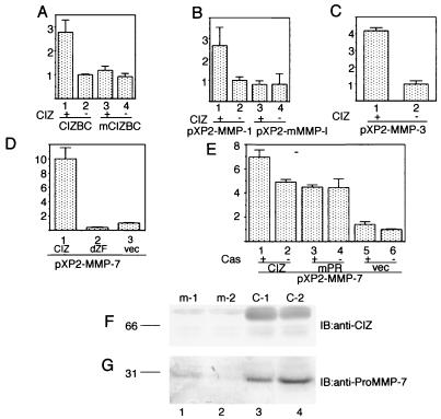
p130(cas) (Cas) is a docking protein that contains an SH3 domain and multiple tyrosine residues. p130(cas) is located at focal adhesions, is tyrosine phosphorylated in response to integrin stimulation, and is thought to transmit signals, via c-Crk and other proteins, for the remodeling of actin stress fibers and cell movement. In a search for the ligands of the SH3 domain of p130(cas) by far-Western screening, we cloned a novel protein named CIZ (for Cas-interacting zinc finger protein). CIZ consists of the following: a putative leucine zipper; a serine/threonine-rich region; a proline-rich sequence; five, six, or eight Krüppel-type C(2)H(2) zinc fingers; and the glutamine-alanine repeat. CIZ binds Cas in cells and is located in the nucleus and at focal adhesions. We showed that CIZ is a nucleocytoplasmic shuttling protein, by using the transient interspecies heterokaryon formation assay. In order to search for the targets of CIZ in nucleus, we determined the DNA binding consensus of CIZ as (G/C)AAAAA(A) by cyclic amplification and selection of targets analysis. The consensus-like sequences are found in several promoters of matrix metalloproteinases (MMPs), which are the enzymes used to degrade the extracellular matrix proteins. CIZ binds to a consensus-like sequence in the MMP-1 (collagenase) promoter. Overexpression of CIZ upregulates the transcriptions from MMP-1, MMP-3 (stromelysin), and MMP-7 (matrilysin) promoters, and this transactivation was enhanced in the presence of Cas. Furthermore, the stable overexpression of CIZ promoted the production of MMP-7 in culture medium. In summary, CIZ, a novel zinc finger protein, binds Cas, is a nucleocytoplasmic shuttling protein, and regulates the expression of MMPs.


Figures

FIG. 1
FIG. 1
Nucleotide and predicted amino acid sequences of rat CIZ cDNA. (A) Nucleotide sequences and predicted amino acid sequences (single-letter code) of rat CIZ cDNA are presented. Shown in lowercase letters are noncoding regions. Shown in italic type are the leucine or isoleucine residues, which appear every six or seven amino acid residues in the N-terminal portion. Shown in boldface type are the serine residues, which are rich in the following region. Overbarred residues are the proline-rich sequence, which was shown to be the Cas SH3 binding site in this paper. Underlined residues are the eight zinc fingers, which are numbered underneath. The boxed nucleotides are the possible CAG repeat portion of CIZ cDNA. <> indicates the residues that are missing in the alternative forms CIZ6-3 and CIZ5. ∗ indicates the insertion point of the residues shown in Fig. 1B, in the alternative forms CIZ6-1 and CIZ6-3. () indicates the residues that are missing in the alternative form CIZ5. {} indicates the residues that are missing in the alternative forms CIZ 6-1, CIZ6-2, CIZ6-3, and CIZ5. (B) Inserted nucleotide sequence and predicted amino acid sequences, inserted at the ∗ point of panel A, in the alternative forms CIZ6-3 and CIZ5.
FIG. 2
FIG. 2
Alternative splicing forms and tissue distribution of CIZ. (A) Schematic diagram indicating the relationship among the clones obtained, including five alternative forms. LZ, leucine zipper; PR, proline rich; NLS, nuclear localization signal; ZFs, zinc fingers; QA, glutamine-alanine repeat. (B) Immunoblotting (IB) with anti-CIZ of the CIZ alternatives expressed in COS-7 cells and of the native CIZ in 3Y1 cells. The lanes are the lysates of mock (lane 1)-, CIZ8 (lane 2)-, CIZ6-1 (lane 3)-, CIZ6-2 (lane 4)-, CIZ6-3 (lane 5)-, and CIZ5 (lane 6)-transfected COS-7 cells and 3Y1 cell lysate (lane 7). (C) An RNA blot (Clontech) containing poly(A)+ RNA from the indicated rat tissues (2 μg/lane) was hybridized with a labeled CIZ partial cDNA probe (RsaI fragment corresponding to 234 to 481 bp). The positions of 9.5-, 7.5-, 4.4-, 2.4-, and 1.35-kb markers are shown on the left. (D) As a control, a blot with the cDNA of human β-actin is shown.
FIG. 3
FIG. 3
Association of CIZ with Cas. (A) FLAG-tagged CIZ expressed in COS-7 cells was precipitated with GST (lane 1), GST-Cas SH3 (lane 2), GST-Src SH3 (lane 3), GST-Fyn SH3 (lane 4), GST-Crk SH3 (lane 5), GST-Ash/Grb2 SH3 (lane 6), and GST-Abl SH3 (lane 7) and was immunoblotted with anti-FLAG (upper row). The samples were electrophoresed and stained with Coomassie blue (bottom row). Total lysates of COS-7 cells expressing CIZ-FLAG were also immunoblotted (lane 8). (B) FLAG-tagged CIZ (lanes 1 and 5), mPR mutant of CIZ-FLAG (lanes 2 and 6), and mock (lanes 3 and 7)-transfected COS-7 cell lysates were incubated with GST-Cas SH3 (lanes 1, 2, and 3) or with GST (lanes 5, 6, and 7) and immunoblotted with anti-FLAG. Lanes 4 and 8 contained GST-Cas SH3 or GST alone, respectively. vec, vector. (C) CIZ-FLAG was expressed in COS-7 cells, and the lysates were immunoprecipitated (IP) with anti-Cas2 (lane 1) or with preimmune (Pre) serum (lane 2). The total cellular lysates (TL) are shown in lane 3. The samples were immunoblotted with anti-Cas2 in the upper row and immunoblotted with anti-FLAG in the bottom row. (D) Association between CIZ and Cas expressed in COS-7 cells. CIZ-FLAG (lanes 2 and 6) or mPR-FLAG (lanes 1 and 5) was expressed in COS-7 cells together with Cas. Cas alone (lanes 3 and 7) and CIZ-FLAG alone (lanes 4 and 8) were also expressed. In lanes 1 to 4, the lysates were immunoprecipitated with anti-FLAG. In the upper row, the filter was immunoblotted with anti-Cas2. In the bottom row, the same immunoprecipitant was immunoblotted with anti-FLAG. (E) The wild-type Cas (wtCas) (lanes 1 and 3) and the mutant of Cas that lacks the SH3 domain (lanes 2 and 4 [dSH3]) were expressed together with FLAG-tagged CIZ. The lysates were immunoprecipitated with anti-FLAG antibody and immunoblotted with anti-Cas2 (upper row) or anti-CIZ (lower row). (F) The lysates of LC-540 cells were immunoprecipitated with preimmune serum (lane 1), anti-CIZ (lane 2), and anti-Cas2 (lane 3). In the upper row, the filter was immunoblotted with anti-Cas2, and in the lower row, the filter was immunoblotted with anti-CIZ.
FIG. 4
FIG. 4
Subcellular localization of CIZ. (A to F) 3Y1 cells were grown overnight on the uncoated coverslips. (G and H) After overnight culture, 3Y1 cells were treated with LMB (10 ng/ml) for 24 h. Immunofluorescence was performed with anti-CIZ (1:400) (A, C, and G) precleared by GST or with anti-CIZ precleared by GST-CIZ (E), together with antivinculin antibody (B, D, F, and H) as described previously (31). Panels A, B, G, and H show the sections at the height of focal adhesions. Panels C, D, E, and F show the sections at the height of the nucleus.
FIG. 5
FIG. 5
Nuclear-cytoplasmic shuttling of CIZ. (A) Nontransfected HeLa cells were stained with Hoechst 33258 dye. (B) The same cells as in panel A stained with anti-HA. (C) Nontransfected NIH 3T3 cells were stained with Hoechst 33258. (D) The same cells as in panel C stained with anti-HA. (E) A heterokaryon of HeLa cells, CIZ-HA-transfected NIH 3T3 cells, and a nearby NIH 3T3 cell were stained with Hoechst 33258. (F) The same cells as in panel E stained with anti-HA.
FIG. 6
FIG. 6
CIZ has DNA-binding consensus sequences. (A) Results of CASTing described in Materials and Methods. (B) Overexpressed CIZ binds to the CCTTTTTCAAAAAGA sequence of the human MMP-1 promoter. EMSA was performed with the nuclear extract of CIZ-FLAG (lanes 2 to 5)-transfected cells and with that of mock (lane 1)-transfected COS-7 cells. The horizontally lined arrowhead indicates the DNA-protein complex (lanes 2, 4, and 6). The supershifted band is indicated by the vertically lined arrowhead (lane 5). The background band caused by immunoglobulin was indicated by the open arrowhead (lanes 5 and 6). (C) Endogeneous CIZ is included in the DNA-protein complex (lane 7). The supershift band was observed in panel B (lane 10).
FIG. 7
FIG. 7
CIZ upregulates the expression of MMPs. (A) CIZ transactivates the artificial promoter consisting of four tandem repeats of CIZ-binding consensus sequences in the human MMP-1 promoter. (B to D) CIZ upregulates the transcription from the human MMP-1 promoter mediated by the CIZ-binding consensus sequence (B) from the MMP-3 promoter (C) and from the MMP-7 promoter (D). vec, vector. The effect of Cas expression on the transactivation by CIZ was checked in Cas−/− fibroblasts (E). In these experiments, NIH 3T3 cells or Cas−/− fibroblasts were transfected with each expression vector, together with the reporter construct and β-galactosidase expression vector. Values are the means of three experiments, with bars representing the standard deviations. (F and G) Overproduction of MMP-7 in HT1080 cells stably expressing CIZ. (F) Total lysates of m-1 (lane 1), m-2 (lane 2), C-1 (lane-3), and C-2 (lane-4) cells were immunoblotted (IB) with anti-CIZ. (G) The conditioned media of m-1 (lane 1), m-2 (lane 2), C-1 (lane 3), and C-2 (lane 4) cells were immunoblotted with anti-proMMP-7.

References

    1. Aggeler J, Frisch S M, Werb Z. Changes in cell shape correlate with collagenase gene expression in rabbit synovial fibroblasts. J Cell Biol. 1984;98:1662–1671. - PMC - PubMed
    1. Angel P, Baumann I, Stein B, Delius H, Rahmsdorf H J, Herrlich P. 12-O-Tetradecanoyl-phorbol-13-acetate induction of the human collagenase gene is mediated by an inducible enhancer element located in the 5′-flanking region. Mol Cell Biol. 1987;7:2256–2266. - PMC - PubMed
    1. Astier A, Avraham H, Manie S N, Groopman J, Canty T, Avraham S, Freedman A S. The related adhesion focal tyrosine kinase is tyrosine-phosphorylated after beta1-integrin stimulation in B cells and binds to p130cas. J Biol Chem. 1997;272:228–232. - PubMed
    1. Borer R A, Lehner C F, Eppenberger H M, Nigg E A. Major nucleolar proteins shuttle between nucleus and cytoplasm. Cell. 1989;56:379–390. - PubMed
    1. Cary L A, Han D C, Polte T R, Hanks S K, Guan J L. Identification of p130Cas as a mediator of focal adhesion kinase-promoted cell migration. J Cell Biol. 1998;140:211–221. - PMC - PubMed

Publication types

MeSH terms

LinkOut - more resources