. 2000 Mar;20(5):1877-85.
doi: 10.1128/MCB.20.5.1877-1885.2000.

The BIR motifs mediate dominant interference and oligomerization of inhibitor of apoptosis Op-IAP

Affiliations

The BIR motifs mediate dominant interference and oligomerization of inhibitor of apoptosis Op-IAP

R R Hozak et al. Mol Cell Biol. 2000 Mar.

Abstract

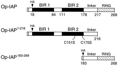
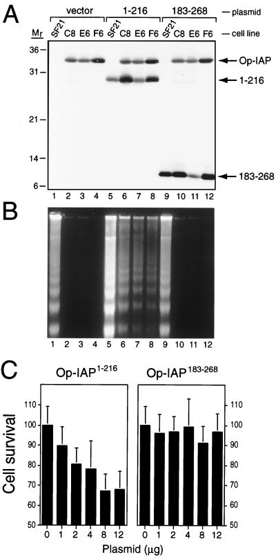
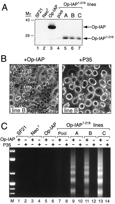
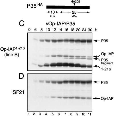
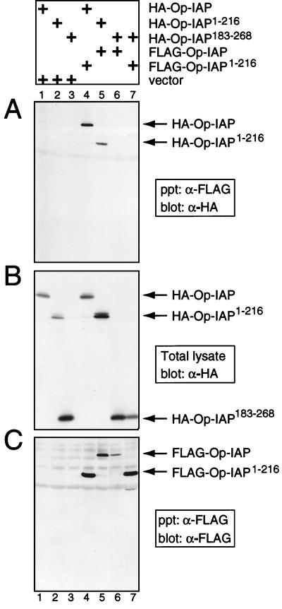
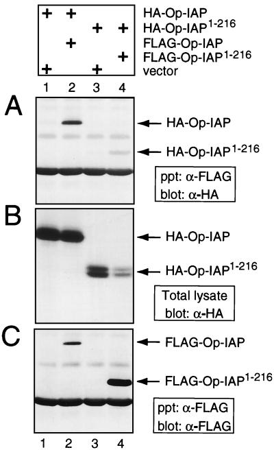
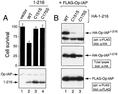
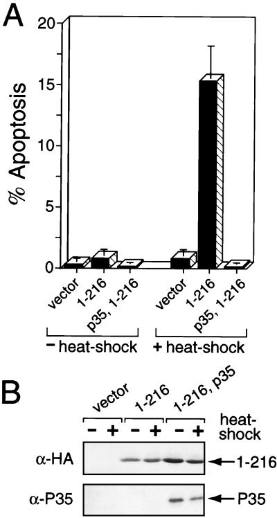
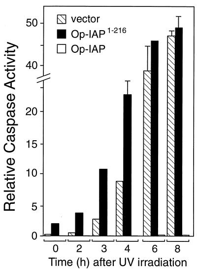
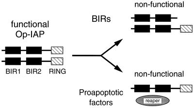
The defining structural motif of the inhibitor of apoptosis (iap) protein family is the BIR (baculovirus iap repeat), a highly conserved zinc coordination domain of approximately 70 residues. Although the BIR is required for inhibitor-of-apoptosis (IAP) function, including caspase inhibition, its molecular role in antiapoptotic activity in vivo is unknown. To define the function of the BIRs, we investigated the activity of these structural motifs within Op-IAP, an efficient, virus-derived IAP. We report here that Op-IAP(1-216), a loss-of-function truncation which contains two BIRs but lacks the C-terminal RING motif, potently interfered with Op-IAP's capacity to block apoptosis induced by diverse stimuli. In contrast, Op-IAP(1-216) had no effect on apoptotic suppression by caspase inhibitor P35. Consistent with a mechanism of dominant inhibition that involves direct interaction between Op-IAP(1-216) and full-length Op-IAP, both proteins formed an immunoprecipitable complex in vivo. Op-IAP also self-associated. In contrast, the RING motif-containing truncation Op-IAP(183-268) failed to interact with or interfere with Op-IAP function. Substitution of conserved residues within BIR 2 caused loss of dominant inhibition by Op-IAP(1-216) and coincided with loss of interaction with Op-IAP. Thus, residues encompassing the BIRs mediate dominant inhibition and oligomerization of Op-IAP. Consistent with dominant interference by interaction with an endogenous cellular IAP, Op-IAP(1-216) also lowered the survival threshold of cultured insect cells. Taken together, these data suggest a new model wherein the antiapoptotic function of IAP requires homo-oligomerization, which in turn mediates specific interactions with cellular apoptotic effectors.


Figures

FIG. 1
FIG. 1
Structures of Op-IAP and truncations. Op-IAP (268 amino acids) contains two BIRs (black boxes) and a C-terminal RING finger motif (crosshatched). Truncation Op-IAP1–216 (residues 1 to 216) contains BIRs 1 and 2 plus the linker domain but lacks the RING finger. Truncation Op-IAP183–268 (residues 183 to 268) contains the linker and RING finger. Each protein contains Op-IAP residues 1 to 5 followed by the influenza virus HA epitope tag. Residues at the boundary of each motif are those previously described (18).
FIG. 2
FIG. 2
Op-IAP1–216 interference with Op-IAP-mediated suppression of UV-induced apoptosis. (A) Protein levels. Parental SF21 cells or stable SF21 lines C8, E6, and F6 that produce full-length Op-IAP were transfected with the vector alone (vector) or a plasmid encoding Op-IAPHA/1–216 or Op-IAPHA/183–268, respectively. Total cell lysates were prepared 18 h after transfection and subjected to immunoblot analysis by using HA-specific antiserum. Proteins Op-IAPHA, Op-IAPHA/1–216 (1–216), and Op-IAPHA/183–268 (183–268) and molecular mass (Mr) markers (sizes are in kilodaltons) are indicated. (B) DNA fragmentation. The cells were transfected with plasmids as indicated in panel A and UV irradiated 18 h later. Low-molecular-weight DNA was collected from cells and apoptotic bodies 12 h after irradiation and subjected to agarose gel electrophoresis in the presence of ethidium bromide. (C) Dose responses. Op-IAP-producing cell line F6 was transfected with increasing amounts of a plasmid encoding Op-IAPHA/1–216 (left) or Op-IAPHA/183–268 (right). A constant level of DNA was maintained by using the vector plasmid. Cells were UV irradiated 19 h after transfection. Cell survival was scored 26 h later and is reported as the average ± the standard deviation of viable cells relative to that of vector-transfected cells (normalized to 100). On the basis of reporter gene (lacZ) expression, about 50% of the SF21 population is transfected under these conditions (data not shown).
FIG. 3
FIG. 3
Effect of Op-IAP1–216 in stably transfected cell lines. (A) Op-IAP1–216 protein levels. Total cell lysates (2.5 × 10⁵ cell equivalents) from pooled (lane 4) or cloned lines A, B, and C (lanes 5 to 7) stably transfected with Op-iapHA/1–216 were subjected to immunoblot analysis by using anti-HA serum. Lysates from parental SF21 (lane 1), Neor (lane 2), and Op-IAP line E6 (lane 3) cells were included. Molecular mass (Mr) markers (sizes are in kilodaltons) are indicated on the left. (B) Apoptosis by dominant inhibition of Op-IAP. Op-IAP1–216 cell line B was inoculated (MOI of 5) with A. californica nucleopolyhedrovirus recombinant vOp-IAP (+Op-IAP) or wt/lacZ (+P35) expressing either Op-iap or p35, respectively. Cells and apoptotic bodies were photographed 18 h later (original magnification, ×100). (C) DNA fragmentation. Pooled or cloned Op-IAP1–216 cell lines, along with SF21, Neor, and Op-IAP cells, were inoculated (MOI of 5) with virus vOp-IAP (Op-IAP) or wt/lacZ (P35). Low-molecular-weight DNA was quantified 24 h later as described in the legend to Fig. 2. DNA molecular weight markers (lane M) were included.
FIG. 4
FIG. 4
Stability of full-length Op-IAP during apoptosis. Op-IAP1–216 cell line B and parental SF21 cells were inoculated (MOI of 5) with virus vOp-IAP (A and B) or vOp-IAP/P35 (C and D). At the indicated times after infection, intact cells and associated apoptotic bodies were collected, lysed with SDS, and subjected to immunoblot analysis (2.5 × 10⁵ cell equivalents) by using anti-HA serum. Proteins Op-IAPHA, Op-IAPHA/1–216 (1–216), and P35HA and the 25-kDa P35HA cleavage fragment are indicated at the right. The cleavage site and the position of the HA epitope within P35 encoded by vOp-IAP/P35 are shown.
FIG. 4
FIG. 4
Stability of full-length Op-IAP during apoptosis. Op-IAP1–216 cell line B and parental SF21 cells were inoculated (MOI of 5) with virus vOp-IAP (A and B) or vOp-IAP/P35 (C and D). At the indicated times after infection, intact cells and associated apoptotic bodies were collected, lysed with SDS, and subjected to immunoblot analysis (2.5 × 10⁵ cell equivalents) by using anti-HA serum. Proteins Op-IAPHA, Op-IAPHA/1–216 (1–216), and P35HA and the 25-kDa P35HA cleavage fragment are indicated at the right. The cleavage site and the position of the HA epitope within P35 encoded by vOp-IAP/P35 are shown.
FIG. 5
FIG. 5
Immunoprecipitation of Op-IAP and Op-IAP1–216. SF21 cells were transfected with plasmids (+) encoding the indicated epitope-tagged proteins or the vector alone, and extracts were prepared ∼24 h later. Proteins (106 cell equivalents) were immunoprecipitated (ppt) by using a FLAG-specific monoclonal antibody (α-FLAG) and subjected to immunoblot analysis by using either anti-HA (A) or anti-FLAG (C) serum. HA-tagged proteins in extracts (2.5 × 10⁵ cell equivalents) prior to immunoprecipitation were quantified by immunoblot analysis using anti-HA serum (B).
FIG. 6
FIG. 6
Intracellular oligomerization of Op-IAP. Immunoprecipitations of extracts from SF21 cells transfected with plasmids (+) encoding the indicated proteins or the vector alone were performed by using anti-FLAG (α-FLAG) serum as described in the legend to Fig. 5. The resulting complexes were subjected to immunoblot analysis with anti-HA (α-HA) (A) or anti-FLAG (C) serum. HA-tagged proteins in extracts (2.5 × 10⁵ cell equivalents) prior to immunoprecipitation were quantified by immunoblot analysis using α-HA (B).
FIG. 7
FIG. 7
Loss of dominant inhibition and interaction by Op-IAP1–216 mutations. (A) Dominant-inhibition assays. SF21 cells were transfected in duplicate with the vector alone or a plasmid encoding WT Op-IAPHA/1–216 or C151S- or C175S-mutated Op-IAPHA/1–216 and infected with vOp-IAP (MOI of 10) 18 h later. Cell survival was scored 24 h after infection and is reported as averages (± the standard deviation) relative to that of vector-transfected cells (normalized to 100). Cell lysates (2.5 × 10⁵ cell equivalents) prepared 7.5 h after infection were subjected to immunoblot analysis by using anti-HA (α-HA) serum (lower panel). (B) Immunoprecipitations. SF21 cells were transfected with a plasmid encoding FLAG-tagged Op-IAP and WT or C151S- or C175S-mutated Op-IAPHA/1–216. Cell extracts prepared 16 h later were immunoprecipitated with anti-FLAG (α-FLAG) serum. The resulting complexes were subjected to immunoblot analysis with anti-HA (top panel) or anti-FLAG (bottom panel) serum. HA-tagged proteins present prior to immunoprecipitation were quantified by using anti-HA serum (central panel).
FIG. 8
FIG. 8
Stress-induced apoptosis by Op-IAP1–216. (A) Transient transfections. SF21 cells were transfected with the vector alone (vector) or a plasmid encoding Op-IAPHA/1–216 (1–216) or P35 (p35). After 18 h, transfected cells were incubated for 30 min at either 27°C (−heat shock) or 42°C (+heat shock) and scored 2.5 to 3 h later for apoptosis by microscopic visualization of cellular membrane blebbing. Reported values are averages ± the 95% confidence intervals of the percent apoptosis for a representative experiment. (B) Protein levels. Transfected cell lysates prepared 2 h after heat shock were subjected to immunoblot analysis by using anti-HA (α-HA) serum.
FIG. 9
FIG. 9
Op-IAP1–216 acceleration of caspase activation. SF21 cells were UV irradiated 18 h after transfection with a plasmid encoding Op-IAPHA/1–216 (solid bars) or the vector alone (striped bars). Cell extracts prepared immediately prior to irradiation (0 h) or at the indicated times after irradiation were assayed for caspase activity by using Ac-DEVD-AMC as the substrate. Relative caspase activities are averages ± the standard deviations of triplicate assays. Caspase activity in extracts from stably transfected Op-IAP cell line E6 after UV irradiation (open bars) was included.
FIG. 10
FIG. 10
Model for dominant inhibition of oligomeric Op-IAP. Op-IAP exhibits antiapoptotic activity as an oligomeric complex through undefined interactions with downstream apoptotic effectors. The BIR-containing truncation Op-IAP1–216 dominantly interferes with Op-IAP by direct interaction, forming an inactive heterocomplex that lowers the survival threshold. By a similar mechanism, proapoptotic factors (invertebrate Reaper, Hid, and Grim) may interact with Op-IAP to form an inactive complex. The balance between proapoptotic factors (determined by the strength of the inductive signal) and endogenous IAP determines cell fate. During infection, the baculovirus prolongs cell survival by overproduction of Op-IAP, thereby overwhelming the death factor activity.

Similar articles

Cited by

References

    1. Adida C, Berrebi D, Peuchmaur M, Reyes-Mugica M, Altieri D C. Anti-apoptotis gene, survivin, and prognosis of neuroblastoma. Lancet. 1998;351:882–883. - PubMed
    1. Ambrosini G, Adida C, Altieri D C. A novel anti-apoptosis gene, survivin, expressed in cancer and lymphoma. Nat Med. 1997;3:917–921. - PubMed
    1. Bergmann A, Agapite J, Steller H. Mechanisms and control of programmed cell death in invertebrates. Oncogene. 1998;17:3215–3223. - PubMed
    1. Bertin J, Mendrysa S M, LaCount D J, Gaur S, Krebs J F, Armstrong R C, Tomaselli K J, Friesen P D. Apoptotic suppression by baculovirus P35 involves cleavage by and inhibition of a virus-induced CED-3/ICE-like protease. J Virol. 1996;70:6251–6259. - PMC - PubMed
    1. Birnbaum M J, Clem R J, Miller L K. An apoptosis-inhibiting gene from a nuclear polyhedrosis virus encoding a polypeptide with Cys/His sequence motifs. J Virol. 1994;68:2521–2528. - PMC - PubMed

Publication types

Substances