. 2000 Mar;20(5):1886-96.
doi: 10.1128/MCB.20.5.1886-1896.2000.

Posttranslational modification of Bcl-2 facilitates its proteasome-dependent degradation: molecular characterization of the involved signaling pathway

Affiliations

Posttranslational modification of Bcl-2 facilitates its proteasome-dependent degradation: molecular characterization of the involved signaling pathway

K Breitschopf et al. Mol Cell Biol. 2000 Mar.

Abstract

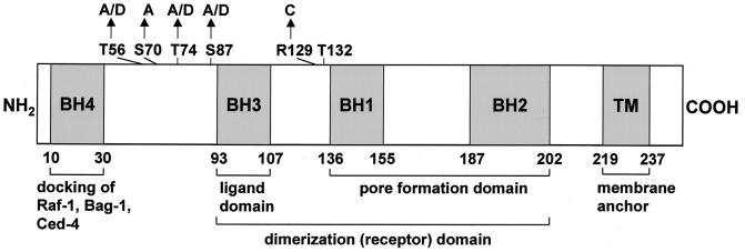
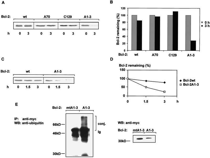
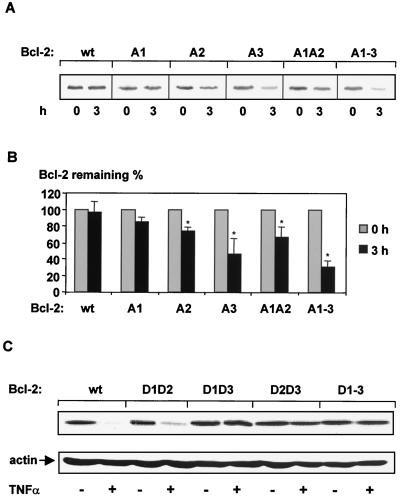
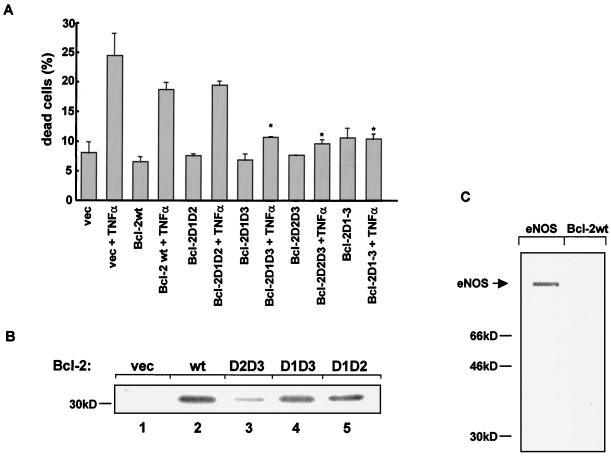
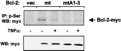
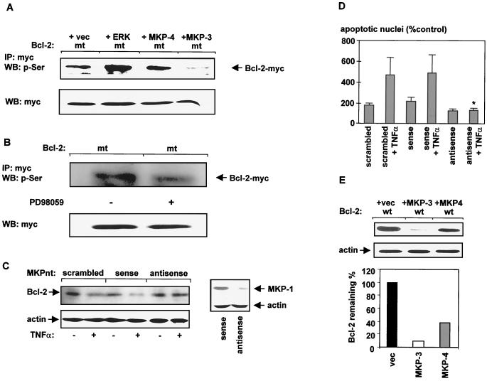
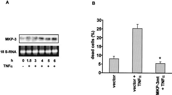
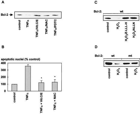
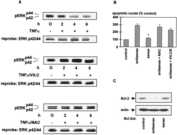
The ratio of proapoptotic versus antiapoptotic Bcl-2 members is a critical determinant that plays a significant role in altering susceptibility to apoptosis. Therefore, a reduction of antiapoptotic protein levels in response to proximal signal transduction events may switch on the apoptotic pathway. In endothelial cells, tumor necrosis factor alpha (TNF-alpha) induces dephosphorylation and subsequent ubiquitin-dependent degradation of the antiapoptotic protein Bcl-2. Here, we investigate the role of different putative phosphorylation sites to facilitate Bcl-2 degradation. Mutation of the consensus protein kinase B/Akt site or of potential protein kinase C or cyclic AMP-dependent protein kinase sites does not affect Bcl-2 stability. In contrast, inactivation of the three consensus mitogen-activated protein (MAP) kinase sites leads to a Bcl-2 protein that is ubiquitinated and subsequently degraded by the 26S proteasome. Inactivation of these sites within Bcl-2 revealed that dephosphorylation of Ser87 appears to play a major role. A Ser-to-Ala substitution at this position results in 50% degradation, whereas replacement of Thr74 with Ala leads to 25% degradation, as assessed by pulse-chase studies. We further demonstrated that incubation with TNF-alpha induces dephosphorylation of Ser87 of Bcl-2 in intact cells. Furthermore, MAP kinase triggers phosphorylation of Bcl-2, whereas a reduction in Bcl-2 phosphorylation was observed in the presence of MAP kinase-specific phosphatases or the MAP kinase-specific inhibitor PD98059. Moreover, we show that oxidative stress mediates TNF-alpha-stimulated proteolytic degradation of Bcl-2 by reducing MAP kinase activity. Taken together, these results demonstrate a direct protective role for Bcl-2 phosphorylation by MAP kinase against apoptotic challenges to endothelial cells and other cells.


Figures

FIG. 1
FIG. 1
Schematic diagram of Bcl-2 (1). Different homologue domains (BH) are indicated by hatched boxes. Putative phosphorylation sites investigated in this study are indicated. MAP kinase sites, T56, T74, and S87; PKC or PKA sites, S70; Akt kinase site, T132. Arrows indicate substitutions. A, alanine; C, cysteine; D, aspartic acid; S, serine; T, threonine; TM, transmembrane domain.
FIG. 2
FIG. 2
Stability of various Bcl-2 constructs with different inactivated phosphorylation sites. (A) Bcl-2 stability is affected neither by the evolutionarily conserved PKA or PKC site Ser70 nor by the Akt site Thr132. Bcl-2 proteins metabolically labeled with 35S were chased (3 h) as described in Materials and Methods. Labeled Bcl-2 was immunoprecipitated from aliquots containing equal amounts of proteins. Bcl-2wt, wild-type Bcl-2; Bcl-2A70, Ser-to-Ala substitution at position 70; Bcl-2C129, destruction of the Akt consensus site by an exchange of Arg with Cys; Bcl-2A1-3, MAP kinase consensus sites Thr56, Thr74, and Ser87 were changed to Ala. A representative autoradiogram of three independent experiments is shown. (B) Quantitative (PhosphorImaging) analysis of the data depicted in panel A is shown. Quantities are relative to the amount of protein at time zero. (C) Representative time course degradation of wild-type Bcl-2 (wt) and of a Bcl-2 construct in which all MAP kinase consensus sites were changed to Ala (Bcl-2A1-3) (n = 3). (D) Quantitative analysis of the data depicted in panel C. Quantities are relative to the amount of protein at time zero. (E) Detection of ubiquitin-Bcl-2 conjugates in HeLa cells. HeLa cells were transiently transfected with expression vector containing either a lysine-free Bcl-2A1-3 construct (Bcl-2mtA1-A3) or Bcl-2A1-3. Following 42 h of transfection, cells were incubated for an additional 2 h with the proteasome inhibitor lactacystin. Equal amounts of protein, as determined by the Bradford method (6), were subjected to immunoprecipitation with anti-myc antibody, and ubiquitin conjugates were identified by Western blot (WB) analysis and anti-ubiquitin antibody. Expression of Bcl-2 protein is detected by Western blot analysis with anti-myc antibody. conj., conjugates; Ig, the heavy chain of the immunoglobulin molecule.
FIG. 3
FIG. 3
In vivo degradation of Bcl-2 mutated proteins in which the MAP kinase sites were progressively substituted with Ala. (A) HeLa cells transiently transfected with Bcl-2 cDNA were pulse labeled (2 h) with [35S]methionine and chased (3 h) as described in Materials and Methods. Bcl-2A1, Thr56 was replaced with Ala; Bcl-2A2, Thr74 was changed to Ala; Bcl-2A3, Ser87 was mutated to Ala; Bcl-2A1A2, combined substitution of Thr56 and Thr74 with Ala; Bcl-2A1-A3, Thr56, Thr74, and Ser87 were replaced with Ala. A representative autoradiogram is shown (n = 3). (B) Quantitative analysis of three independent experiments described for panel A after a chase period of 3 h. Quantities are relative to the amount of protein at time zero. Data are mean ± SEM (error bars) (∗, significantly different from amount of wild-type Bcl-2 protein after a 3-h chase [P < 0.05], n = 3). (C) Sensitivity of various Bcl-2 constructs containing phosphate-mimetic amino acids at relevant MAP kinase sites to TNF-α. HUVEC transiently transfected with Bcl-2 in pcDNA3.1 were incubated with TNF-α (100 ng/ml) for 6 h. Bcl-2D1D2, Thr56 and Thr74 were replaced with Asp; Bcl-2D1D3, Thr56 and Ser87 were replaced with Asp; Bcl-2D2D3, Thr74 and Ser87 were changed to Asp; Bcl-2D1-D3, Thr56, Thr74, and Ser87 were mutated to Asp. Western blot analysis was carried out with anti-myc antibody. Following stripping of the PVDF membrane, equal loading of the samples was demonstrated by Western blot analysis with antiactin antibody.
FIG. 4
FIG. 4
Influence of phosphate-mimetic Bcl-2 mutant proteins on apoptosis and in vitro kinase assays of various Bcl-2 proteins. (A) HUVEC were transiently cotransfected with a vector carrying either wild-type Bcl-2 or various phosphate-mimetic Bcl-2 constructs and a lacZ reporter. Apoptosis was induced by incubation with TNF-α (100 ng/ml) for 18 h. Transfected cells were identified by β-galactosidase staining as described under Materials and Methods. Data are mean + SEM (error bars) (∗, significantly different from Bcl-2wt + TNF-α [P < 0.05]; n = 4). vec, empty vector. (B) Phosphorylation of various Bcl-2 constructs by active MAP kinase. Wild-type Bcl-2 or mutant Bcl-2 forms lacking two of the three putative MAP kinase acceptor amino acids were expressed in HeLa cells and myc-tagged Bcl-2 was immunoprecipitated with anti-myc antibody. Isolated immunocomplexes were incubated with active MAP kinase as described under Materials and Methods and resolved by SDS-PAGE. Lane 1, empty vector (vec); lane 2, wild type (wt); lane 3, Bcl-2D2D3 (Thr74 and Ser87 were changed to Asp); lane 4, Bcl-2D1D3 (Thr56 and Ser87 are with Asp); lane 5, Bcl-2D1D2 (Thr56 and Thr74 were replaced by Asp). (C) In vitro phosphorylation of Bcl-2wt and endothelial NO synthase (eNOS) by constitutive active kinase Akt. The kinase assay was carried out as described in Materials and Methods. Experiments were repeated three times, with identical results.
FIG. 5
FIG. 5
Effect of TNF-α on Ser87 phosphorylation of Bcl-2. HUVEC were transfected with empty vector (vec), lysine-free myc-tagged Bcl-2 (Bcl-2mt [Lys17Arg, Lys22Arg, Lys218Arg, and Lys239Arg]), or lysine-free Bcl-2A1-3 (Bcl-2mtA1-3 [Thr56Ala, Thr74Ala, Ser87Ala, Lys17Arg, Lys22Arg, Lys218Arg, and Lys239Arg]) and incubated with or without TNF-α (100 ng/ml) for 6 h. Serine-phosphorylated Bcl-2 was immunoprecipitated (IP) with antiphosphoserine antibody. Bcl-2 protein levels were detected by Western blot (WB) analysis with anti-myc antibodies. Western blot of cell lysates with antibody against myc (lower panel) served as a control for expression. A representative blot of three independent experiments is shown.
FIG. 6
FIG. 6
ERK2 induces Bcl-2 phosphorylation whereas ERK-specific phosphatases induce Bcl-2 dephosphorylation and its subsequent degradation. (A) Plasmids encoding a myc-tagged lysine-free Bcl-2 protein (Bcl-2mt) were cotransfected with either MKP-3, MKP-4, ERK in pcDNA3.1, or empty vector (vec) in HeLa cells. After 42 h of transfection, ERK activity was stimulated by starving cells in FCS-free medium for 2 h and a subsequent addition of FCS for 1 h. Immunoprecipitation (IP) was performed with anti-myc antibody. Immunocomplexes were resolved by SDS-PAGE as described under Materials and Methods. Western blot (WB) analysis was carried out with antiphosphoserine antibody. Western blot analysis of protein homogenates with anti-myc antibody served as a control for Bcl-2 expression. (B) Effect of PD98059 on serine phosphorylation. Lysine-free myc-tagged Bcl-2 protein (Bcl-2mt) was transfected into HeLa cells and 30 h after transfection, cells were incubated with PD98059 (15 μM) for 18 h. Proteins were immunoprecipitated with anti-myc antibody, and the presence of phosphorylated Bcl-2 was determined with antiphosphoserine antibody. (C) Effect of antisense MKP oligonucleotides on TNF-α-induced degradation of Bcl-2. HUVEC were transfected with either sense, antisense, or scrambled MKP oligonucleotides (MKPnt) by the Lipofectamine method as described in Materials and Methods and incubated for 6 h with or without TNF-α (100 ng/ml). Suppression of MKP-1 after antisense oligonucleotide treatment is shown in the right panel via Western blot analysis with anti-MKP-1 antibody. Stripping of the PVDF membrane followed by reprobing with antiactin demonstrated equal loading of the samples. (D) TNF-α-induced apoptosis in HUVEC is completely inhibited in the presence of antisense MKP oligonucleotides. Lipofectamine-treated cells served as controls. (∗, significantly different from sense + TNF-α [P < 0.05]; n = 3; mean ± SEM [error bars] are shown; apoptosis in cells transfected with Lipofectamine was about 10%). (E) Effect of MKP-3 and MKP-4 on Bcl-2 stability. HeLa cells were cotransfected with Bcl-2wt and either empty vector (vec), MKP-3, or MKP-4. Forty-two hours after transfection, cells were lysed and proteins were separated by SDS–12.5% PAGE. Western blot analysis was performed with anti-myc antibody. Reprobe of the PVDF membrane with antiactin demonstrated equal loading of the samples (middle panel). The lower panel shows a quantitative analysis of the data depicted in the upper panel. Quantities are relative to the amount of Bcl-2 protein cotransfected with empty vector (Bcl-2 + vec). Each experiment was performed three times, with identical results.
FIG. 7
FIG. 7
Effect of TNF-α on MKP-3 expression. (A) Northern blot analysis of MKP-3 mRNA after stimulation of HUVEC with TNF-α. Equal loading of the samples is demonstrated by determining 18S RNA concentration. (B) TNF-α-induced apoptosis in HUVEC is completely inhibited by overexpression of a dominant negative MKP-3 (MKP-3mt) mutant protein. Data are mean ± SEM (error bars) (∗, significantly different from vector + TNF-α [P < 0.05], n = 3). MKP-3mt, Cys293 was mutated to Ser.
FIG. 8
FIG. 8
Effect of antioxidants on TNF-α-stimulated Bcl-2 degradation and apoptosis in HUVEC. (A) Inhibition of TNF-α-stimulated degradation of Bcl-2 in vivo. HUVEC were incubated for 12 h in the presence of 100 ng of TNF-α/ml and vitamins C and E (Vit.C/E; 10 μM concentrations of each), NAC (200 μM), or pyrrolidinedithiocarbamate (PDTC; 10 μM). Bcl-2 protein levels were determined by Western blotting. (B) HUVEC were incubated with TNF-α (100 ng/ml) in the presence or absence of NAC (200 μM) or vitamins C and E (10 μM) for 18 h, and apoptosis was assessed by morphological analysis of DAPI-stained nuclei (∗, significantly different from TNF-α [P < 0.05]; apoptosis in nontransfected cells was about 1.0%). (C) Effect of ROS on Bcl-2 stability in vivo. HUVEC were incubated with H2O2 (200 μM) for 12 h in the presence or absence of the proteasome inhibitors Z-LLL-H (20 μM) or ALLN (0.5 μg/ml). (D) HUVEC transiently transfected with either a wild-type or a degradation-resistant Bcl-2 construct (Bcl-2mt) were incubated with H2O2 (200 μM) for 12 h. A representative Western blot against myc-tagged Bcl-2 is shown. Each experiment was repeated three times, with identical results.
FIG. 9
FIG. 9
(A) Antioxidants inhibit TNF-α-triggered ERK1/2 deactivation. HUVEC were incubated with TNF-α (100 ng/ml) in the presence of either 100 μM vitamin C (Vit.C) or 200 μM NAC. Cells were lysed, and Western blot analysis was carried out with anti-phospho-ERK1/2 antibody to determine activated, phosphorylated ERK1/2. Following stripping of the PVDF membrane, equal loading of the samples was demonstrated by Western blot analysis with anti-ERK antibody. Experiments were performed three times, with identical results. (B) Effect of antioxidants on apoptosis induced by antisense oligonucleotides against Bcl-2. HeLa cells were transfected with antisense or sense oligonucleotides directed against Bcl-2 for 18 h, and apoptosis was determined by morphological analysis of DAPI-stained nuclei (∗, significantly different from antisense oligonucleotides [P < 0.05]). Experiments were performed three times, with identical results. (C) Effect of antisense Bcl-2 oligonucleotides on Bcl-2 expression. HUVEC were transfected with either sense or antisense Bcl-2 oligonucleotides (Bcl-2nt) by the Lipofectamine method described in Materials and Methods. Suppression of Bcl-2 after antisense oligonucleotide treatment is shown via Western blot analysis with anti-Bcl-2 antibody. Stripping of the PVDF membrane followed by reprobing with antiactin demonstrates equal loading of the samples.

Similar articles

Cited by

References

    1. Adams J M, Cory S. The Bcl-2 protein family: arbiters of cell survival. Science. 1998;281:1322–1325. - PubMed
    1. Arch R H, Gedrich R W, Thompson C B. Tumor necrosis factor receptor-associated factors (TRAFs)—a family of adapter proteins that regulates life and death. Genes Dev. 1998;12:2821–2830. - PubMed
    1. Ashkenazi A, Dixit V M. Death receptors: signaling and modulation. Science. 1998;281:1305–1308. - PubMed
    1. Baffy G, Miyashita T, Williamson J R, Reed J C. Apoptosis induced by withdrawal of interleukin-3 (IL-3) from an IL-3-dependent hemapoietic cell line is associated with repartitioning of intracellular calcium and is blocked by enforced Bcl-2 oncoprotein production. J Biol Chem. 1993;268:6511–6519. - PubMed
    1. Batt D B, Carmichael G G, Liu Z. An improved rapid method of isolating RNA from cultured cells. Methods Mol Biol. 1998;86:15–17. - PubMed

Publication types

MeSH terms

Substances

LinkOut - more resources