

Review
doi: 10.1128/JB.182.5.1191-1199.2000.
Cyanobacterial cell walls: news from an unusual prokaryotic envelope
Affiliations
- PMID: 10671437
- PMCID: PMC94402
- DOI: 10.1128/JB.182.5.1191-1199.2000
Item in Clipboard
Review
Cyanobacterial cell walls: news from an unusual prokaryotic envelope
J Bacteriol.
2000 Mar.
No abstract available
Figures
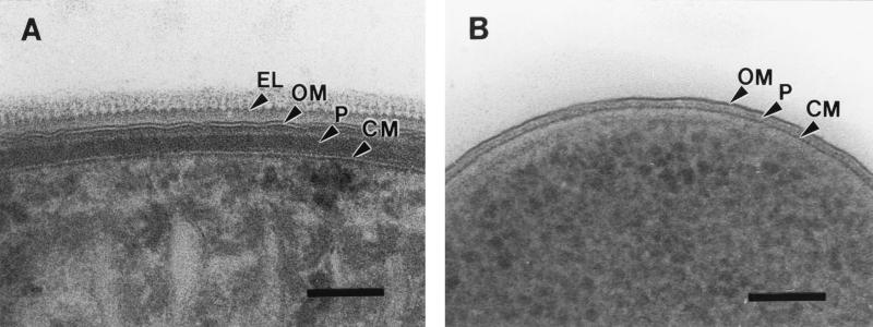
Electron microscopical comparison of the gram-negative cell envelopes of the cyanobacterium P. uncinatum (A) and E. coli (B). Both bacteria were identically processed using cryosubstitution procedures. Note the combination of gram-positive and gram-negative features present in the cyanobacterial cell wall, like the thick peptidoglycan layer and the outer membrane, respectively. The external layer of Phormidium is composed of an S-layer (see Fig. 3) and oscillin fibrils creating a serrated surface topography. CM, cytoplasmic membrane; EL, serrated external layer; OM, outer membrane; P, peptidoglycan layer. Bars, 100 nm.
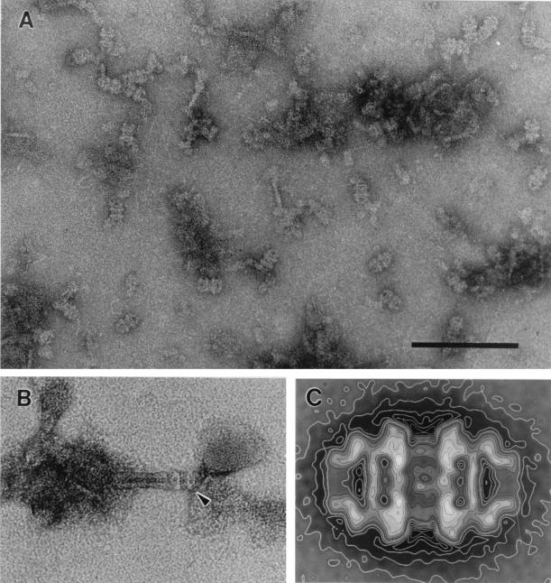
Electron micrographs of negatively stained JPC of P. uncinatum, the carbohydrate secretion organelles which generate the thrust for locomotion in cyanobacteria. (A) Highly enriched fraction of isolated pore complexes. Bar, 150 nm. (B) Appearance of the holo-organelle consisting of a tube-like part and the attached pore complex. (C) Average of side-view projections of the pore complex. The overall length of the complex is 32 nm, measuring 8 nm at the terminal opening. Reproduced from reference , with permission of Elsevier Science.
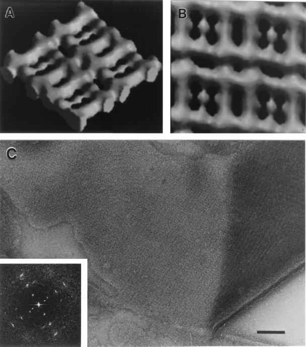
Structure of the oblique S-layer of P. uncinatum (54): Computer-generated side (A) and top (B) view of a surface-shaded, solid three-dimensional model of one side of the S-layer. According to the distribution of the measured data in Fourier space, the resolution of the reconstruction used for the model was about 2.0 nm. The lattice constants of the S-layer are a is 10 nm, b is 9.6 nm, and γ is 97.5°. (C) Appearance of the P. uncinatum cell wall after mechanical preparation of the S-layer (also see reference 56). The inset shows the corresponding optical diffraction pattern (power spectrum) which has been used for the reconstruction of the S-layer.
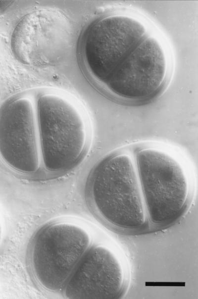
Differential interference contrast micrograph of the sheath of Chroococcus turgidus. This cyanobacterium forms regular aggregates of up to four individual cells which are held together by a multilayered, well-defined sheath. Note that the spherical cells become hemispherical as a consequence of compression applied to the cells by the surrounding sheath. Bar, 10 μm.
References
-
- Archibald A R, Hancock I C, Harwood C R. Cell wall structure, synthesis and turnover. In: Sonenshein A L, Hoch J A, Losick R, editors. Bacillus subtilis and other gram-positive bacteria. Washington, D.C.: American Society for Microbiology; 1993. pp. 381–410.
-
- Baumeister W, Engelhardt H. Three-dimensional structure of bacterial surface layers. In: Harris R, Horne R W, editors. Electron microscopy of proteins. Vol. 6. London, United Kingdom: Academic Press; 1987. pp. 109–154.
-
- Bayer M E. Areas of adhesion between wall and membranes of Escherichia coli. J Gen Microbiol. 1968;53:395–404. - PubMed
Publication types
MeSH terms
LinkOut - more resources
Full Text Sources
Other Literature Sources
