. 2000 Feb 15;523 Pt 1(Pt 1):19-28.
doi: 10.1111/j.1469-7793.2000.t01-1-00019.x.

Reversible Ca2+-induced fast-to-slow transition in primary skeletal muscle culture cells at the mRNA level

Affiliations

Reversible Ca2+-induced fast-to-slow transition in primary skeletal muscle culture cells at the mRNA level

J D Meissner et al. J Physiol. .

Abstract

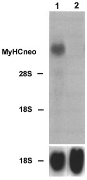
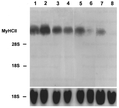
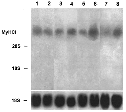
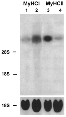
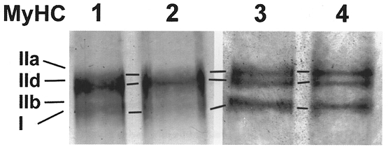
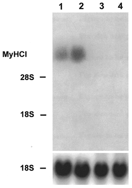
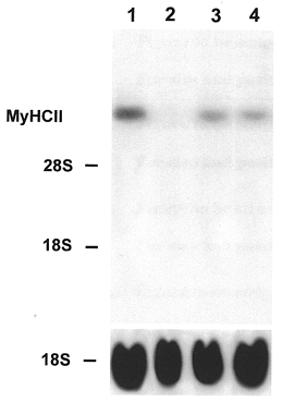
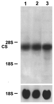
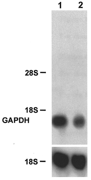
1. The adult fast character and a Ca2+-inducible reversible transition from a fast to a slow type of rabbit myotube in a primary culture were demonstrated at the mRNA level by Northern blot analysis with probes specific for different myosin heavy chain (MyHC) isoforms and enzymes of energy metabolism. 2. No non-adult MyHC isoform mRNA was detected after 22 days of culture. After 4 weeks of culture the fast MyHCIId mRNA was strongly expressed while MyHCI mRNA was virtually absent, indicating the fast adult character of the myotubes in the primary skeletal muscle culture. 3. The data show that a fast-to-slow transition occurred in the myotubes at the level of MyHC isoform gene expression after treatment with the Ca2+ ionophore A23187. The effects of ionophore treatment were decreased levels of fast MyHCII mRNA and an augmented expression of the slow MyHCI gene. Changes in gene expression started very rapidly 1 day after the onset of ionophore treatment. 4. Levels of citrate synthase mRNA increased and levels of glyceraldehyde 3-phosphate dehydrogenase mRNA decreased during ionophore treatment. This points to a shift from anaerobic to oxidative energy metabolism in the primary skeletal muscle culture cells at the level of gene expression. 5. Withdrawal of the Ca2+ ionophore led to a return to increased levels of MyHCII mRNA and decreased levels of MyHCI mRNA, indicating a slow-to-fast transition in the myotubes and the reversibility of the effect of ionophore on MyHC isoform gene expression.


Figures

Figure 1
Figure 1. Northern blot analysis of MyHCneo mRNA
The rabbit primary skeletal muscle culture cells were grown for 11 (lane 1) and 22 days (lane 2). Total RNA (20 μg) was isolated at the time points indicated, fractionated on a 1.2 % agarose-formaldehyde gel, and transferred to nitrocellulose. The blots were hybridized with the 32P-labelled 3′ terminal PstI fragment of perinatal MyHC cDNA (1 × 10⁶ c.p.m. ml−1) or an rDNA probe from 18S rRNA (1 × 10⁶ c.p.m. ml−1). The positions of 18S rRNA (1.9 kb) and 28S rRNA (4.8 kb) on the ethidium bromide-stained gel are indicated.
Figure 2
Figure 2. Effect of late addition of Ca2+ ionophore on the expression of fast MyHCII mRNA
Cell cultures were grown for 8 (lane 1), 16 (lane 2), 23 (lane 3) and 29 days (lanes 5 and 7) in the absence or presence of Ca2+ ionophore A23187 (4 × 10⁻⁷ M) from day 22 of the culture for a further 1 day (lane 4) or 7 days (lanes 6 and 8). Total RNA (20 μg) was isolated from control and ionophore treated cultures on the days indicated, fractionated on a 1.2 % agarose-formaldehyde gel, and transferred to nitrocellulose. Lanes 1–6 were probed with the 32P-labelled 3′ terminal PstI fragment of MyHCIId cDNA (1 × 10⁶ c.p.m. ml−1) or an 18S rDNA probe (1 × 10⁶ c.p.m. ml−1). Lanes 7 and 8 were probed with the 3′ terminal 32P-labelled PstI fragment of MyHCIIb cDNA (1 × 10⁶ c.p.m. ml−1) or an rDNA probe from 18S rRNA (1 × 10⁶ c.p.m. ml−1). The positions of 18S rRNA (1.9 kb) and 28S rRNA (4.8 kb) on the ethidium bromide-stained gel are indicated.
Figure 3
Figure 3. Effect of late addition of Ca2+ ionophore on the expression of slow MyHCI mRNA
Cultures were grown for 8 (lane 1), 16 (lane 2), 23 (lane 3), 24 (lane 5) and 29 days (lane 7) in the absence of ionophore. The other lanes represent cultures grown for 22 days without ionophore and thereafter in the presence of Ca2+ ionophore A23187 (4 × 10⁻⁷ M) for 1 day (lane 4), 2 days (lane 6) or 7 days (lane 8). Total RNA (20 μg) was isolated from control and ionophore treated cultures at the time points indicated, fractionated on a 1.2 % agarose-formaldehyde gel, and transferred to nitrocellulose. The blots were hybridized with the 3′ terminal 32P-labelled HinfI fragment of MyHCI cDNA (1 × 10⁶ c.p.m. ml−1) or an rDNA probe from 18S rRNA (1 × 10⁶ c.p.m. ml−1). The positions of 18S rRNA (1.9 kb) and 28S rRNA (4.8 kb) on the ethidium bromide-stained gel are indicated.
Figure 4
Figure 4. Effect of early addition of Ca2+ ionophore on the expression of slow MyHCI mRNA and fast MyHCII mRNA
Cultures were grown for 24 days without ionophore (lanes 1 and 3) or from day 11 for a further 13 days with Ca2+ ionophore A23187 (4 × 10⁻⁷ M) (lanes 2 and 4). Total RNA (20 μg) was isolated from control and ionophore treated cultures at the time points indicated, fractionated on a 1.2 % agarose-formaldehyde gel, and transferred to nitrocellulose. Lanes 1 and 2 were probed with the 32P-labelled 3′ terminal HinfI fragment of MyHCI cDNA (1 × 10⁶ c.p.m. ml−1) or an 18S rDNA probe (1 × 10⁶ c.p.m. ml−1). Lanes 3 and 4 were probed with the 32P-labelled 3′ terminal PstI fragment of MyHCIId cDNA (1 × 10⁶ c.p.m. ml−1) or an rDNA probe from 18S rRNA (1 × 10⁶ c.p.m. ml−1). The positions of 18S rRNA (1.9 kb) and 28S rRNA (4.8 kb) on the ethidium bromide-stained gel are indicated.
Figure 5
Figure 5. Electrophoresis of MyHC isoforms
Electrophoresis of myosin extracts from myotubes growing on microcarriers. MyHC of the control on day 24 (lane 1) and day 29 (lane 2) and isoform pattern of the Ca2+ ionophore A23187 (4 × 10⁻⁷ M) treated cells after 2 (lane 3) and 7 days (lane 4) of incubation.
Figure 6
Figure 6. Reversibility of the effect of Ca2+ ionophore on the expression of slow MyHCI mRNA
Cells were cultured for 22 (lane 1) or 30 days (lane 3) without ionophore or from day 8 for a further 14 days with Ca2+ ionophore A23187 (4 × 10⁻⁷ M) (lane 2). Other cells were cultured from day 8 to 22 with ionophore and then after withdrawal of ionophore for a further 8 days (lane 4). Total RNA (20 μg) was isolated from control and ionophore treated cultures on the days indicated, fractionated on a 1.2 % agarose-formaldehyde gel, and transferred to nitrocellulose. The blots were hybridized with the 3′ terminal 32P-labelled HinfI fragment of MyHCI cDNA (1 × 10⁶ c.p.m. ml−1) or an rDNA probe from 18S rRNA (1 × 10⁶ c.p.m. ml−1). The positions of 18S rRNA (1.9 kb) and 28S rRNA (4.8 kb) on the ethidium bromide-stained gel are indicated.
Figure 7
Figure 7. Reversibility of the effect of Ca2+ ionophore on the expression of fast MyHCII mRNA
Cells were cultured for 22 (lane 1) or 30 days (lane 3) without ionophore or from day 8 for a further 14 days with Ca2+ ionophore A23187 (4 × 10⁻⁷ M) (lane 2). Other cells were cultured from day 8 to 22 with ionophore and then after withdrawal of ionophore for a further 8 days (lane 4). Total RNA (20 μg) was isolated from control and ionophore treated cultures at the time points indicated, fractionated on a 1.2 % agarose-formaldehyde gel, and transferred to nitrocellulose. The blots were probed with the 32P-labelled 3′ terminal PstI fragment of MyHCIId cDNA (1 × 10⁶ c.p.m. ml−1) or an rDNA probe from 18S rRNA (1 × 10⁶ c.p.m. ml−1). The positions of 18S rRNA (1.9 kb) and 28S rRNA (4.8 kb) on the ethidium bromide-stained gel are indicated.
Figure 8
Figure 8. Northern blot analysis of the citrate synthase (CS) mRNA
Muscle cell cultures were grown for 24 days in the absence (lane 1) of ionophore. Ca2+ ionophore A23187 (4 × 10⁻⁷ M) was added for a further 2 days on day 22 (lane 2) or for a further 13 days on day 11 (lane 3) to the cell cultures. Total RNA (20 μg) was isolated from control and ionophore treated cultures, fractionated on a 1.2 % agarose-formaldehyde gel, and transferred to nitrocellulose. The blots were hybridized with the 32P-labelled 800 bp ClaI-EcoRV fragment of CS cDNA (1 × 10⁶ c.p.m. ml−1) or an rDNA probe from 18S rRNA (1 × 10⁶ c.p.m. ml−1). The positions of 18S rRNA (1.9 kb) and 28S rRNA (4.8 kb) on the ethidium bromide-stained gel are indicated.
Figure 9
Figure 9. Northern blot analysis of the glyceraldehyde 3-phosphate dehydrogenase (GAPDH) mRNA
Muscle cell cultures were grown for 29 days in the absence (lane 1) or for 22 days without and then for further 7 days in the presence of Ca2+ ionophore A23187 (4 × 10⁻⁷ M) (lane 2). Total RNA (20 μg) was isolated from control and ionophore treated cultures, fractionated on a 1.2 % agarose- formaldehyde gel, and transferred to nitrocellulose. The blots were hybridized with a 32P-labelled 1.3 kb PstI fragment encompassing the complete GAPDH cDNA (1 × 10⁶ c.p.m. ml−1) or an rDNA probe from 18S rRNA (1 × 10⁶ c.p.m. ml−1). The positions of 18S rRNA (1.9 kb) and 28S rRNA (4.8 kb) on the ethidium bromide-stained gel are indicated.

References

    1. Aigner S, Gohlsch B, Hämäläinen N, Staron RS, Uber A, Wehrle U, Pette D. Fast myosin heavy chain diversity in skeletal muscles of the rabbit: heavy chain IId, not IIb predominates. European Journal of Biochemistry. 1993;211:367–372. - PubMed
    1. Annex BH, Kraus WE, Dohm GL, Williams RS. Mitochondrial biogenesis in striated muscles: rapid induction of citrate synthase mRNA by nerve stimulation. American Journal of Physiology. 1991;260:C266–270. - PubMed
    1. Brownson C, Isenberg H, Brown W, Salmons S, Edwards Y. Changes in skeletal muscle gene transcription induced by chronic stimulation. Muscle and Nerve. 1988;11:1183–1189. - PubMed
    1. Brownson C, Little P, Jarvis JC, Salmons S. Reciprocal changes in myosin isoform mRNAs of rabbit skeletal muscle in response to the initiation and cessation of chronic electrical stimulation. Muscle and Nerve. 1992a;15:694–700. - PubMed
    1. Brownson C, Little P, Mayne C, Jarvis JC, Salmons S. Reciprocal changes in myosin isoform expression in rabbit fast skeletal muscle resulting from the application and removal of chronic electrical stimulation. Symposium of the Society of Experimental Biology. 1992b;46:301–310. - PubMed

MeSH terms

LinkOut - more resources