. 2000 Feb 15;523 Pt 1(Pt 1):211-22.
doi: 10.1111/j.1469-7793.2000.t01-1-00211.x.

Responses of neurones of the rat suprachiasmatic nucleus to retinal illumination under photopic and scotopic conditions

Affiliations

Responses of neurones of the rat suprachiasmatic nucleus to retinal illumination under photopic and scotopic conditions

N C Aggelopoulos et al. J Physiol. .

Abstract

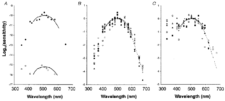
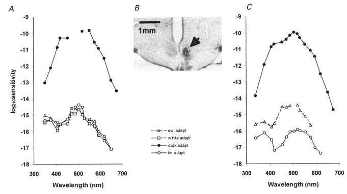
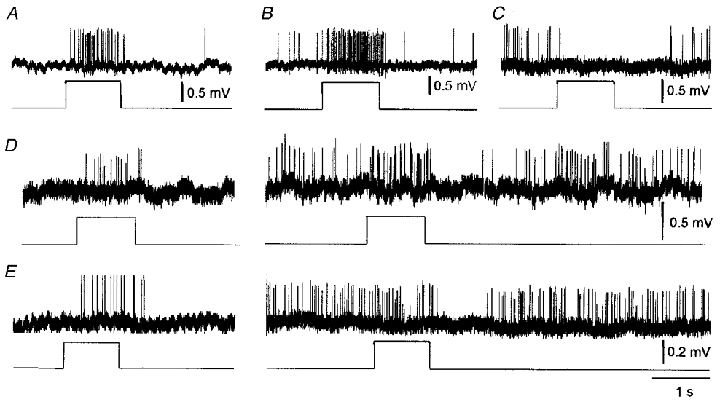
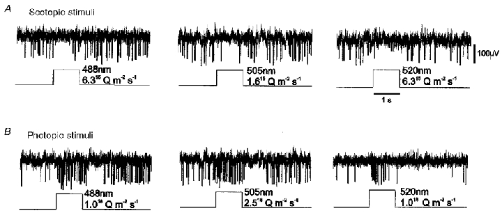
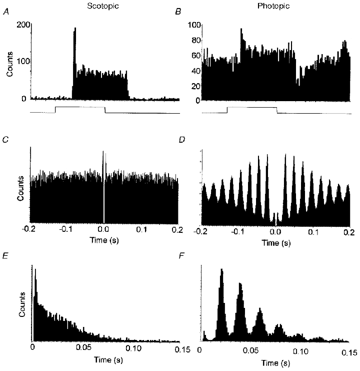
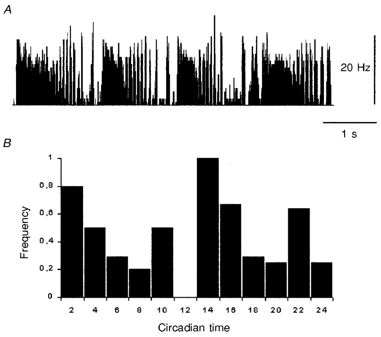
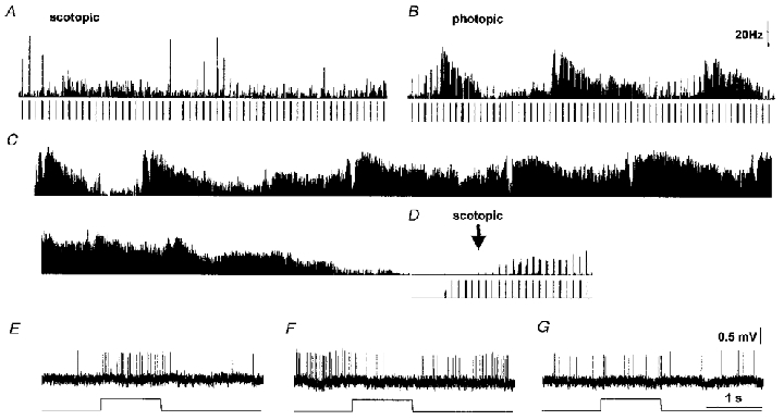
1. We have examined the responses of neurones in the suprachiasmatic nuclei (SCN) of the rat to retinal illumination under photopic and scotopic conditions to identify the types of photoreceptor input to these nuclei. 2. The majority of visually responsive SCN neurones studied under dark adaptation received rod input (48 of 52, 92 %). The action spectrum conformed to the sensitivity of rhodopsin, with maximal sensitivity at around 505 nm. 3. When also studied under light adaptation, most visually responsive SCN neurones (20 out of 26, 77 %) responded to input from cones. The action spectra conformed to the spectrum of green cone opsin, with a main sensitivity peak at 510 nm and a significant secondary peak in the near-ultraviolet region of the spectrum. 4. The frequency of spontaneous activity was typically low under scotopic conditions (range 0.2-17.2 Hz) and higher under photopic conditions (range 0.6-40 Hz) for any given neurone. The most common response under scotopic conditions was an 'on-excitation' (32 of 48, 62.5 %), which changed under photopic conditions to an on-excitation followed by a more prominent off-inhibition. 5. Responses also changed due to endogenous ultradian cycles. Depending on the phase, responses could be altogether absent and even reverted from excitation to inhibition on opposite phases of a cycle. Ultradian cycles had a circadian dependence and were most common at around the light phase:dark phase (L:D) and D:L transition points of the circadian cycle. 6. Under photopic conditions, SCN neurones showed rhythmic electrical activity, with a preferred firing interval that had a value between 18 and 39 ms. This rhythmic activity was probably the result of endogenous subthreshold membrane potential oscillations. 7. In conclusion, light acting either via rod or cone pathways could have powerful, opposing actions on SCN neurones. These actions were state dependent. The presence of these neuronal responses suggests a role for rod and cone photoreceptors in SCN function.


Figures

Figure 1
Figure 1. Action spectra from SCN neurones
A, scotopic and photopic response sensitivity of a neurone in the SCN. Dartnall nomograms have been superimposed on the data points with a peak at 505 nm for the scotopic action spectrum (•) and 510 nm for the photopic action spectrum (○). The threshold for the photopic response is about 5 log units higher than the scotopic response threshold. The Y-axis is logarithmic. The scotopic threshold of about −10 corresponds to 10−10 photons m−2 s−1. B, data points for scotopic sensitivity from 10 different neurones arbitrarily normalised at the 520 nm value. For comparison, the Dartnall nomogram (continuous line, λmax≈505 nm) of the rat rhodopsin and the Lamb equation (dashed line) for the rat rhodopsin have been superimposed on the data points, with the 520 nm value equal to zero. C, data points for photopic sensitivity from 6 different neurones normalised at the 520 nm value. The Dartnall nomogram (continuous line, λmax≈510 nm) for the rat green cone opsin and the Lamb equation (dashed line) for the rat green cone opsin have been superimposed on the data points, with the 520 nm value equal to zero.
Figure 2
Figure 2. Chromatic conditioning of SCN neurones
A, this neurone had a convergence from rods (filled symbols) and cones (open symbols). The cone responses were due to the common type of green cone photoreceptor and had a sensitivity peak at 510 nm with a secondary peak at around 375 nm under white light adaptation (white adapt). Chromatic adaptation with SW light (sw adapt) or LW light (lw adapt) failed to demonstrate independent component mechanisms. The sensitivity curve remained the same under all chromatic adaptation conditions. B, a Nissl-stained coronal section through the SCN showing the location of the recorded unit in A in the right SCN. The injection site (white spot in the lower centre of the nucleus) is surrounded by a silver nitrate sediment (arrow). Compare with the SCN nucleus on the left side. C, another neurone recorded from the SCN with rod- (filled symbols) and cone- (open symbols) mediated responses. The cone response sensitivity peaks could be independently modulated by chromatic adaptation. The green peak was suppressed under LW adaptation, indicating a contribution from another cone photoreceptor with greater sensitivity in the UV region than the typical green cone.
Figure 3
Figure 3. Types of neuronal responses
A, on-excitation. B, on-excitation with both transient and sustained components. C, on-inhibition. D, a neurone with on-excitation under dark adaptation (left) but on-excitation followed by a large off-inhibition under light adaptation (right). E, same as D but with only a transient on-excitatory response remaining under light adaptation, the off-inhibition being the most pronounced effect.
Figure 4
Figure 4. Response reversal in an SCN neurone to light stimuli at scotopic and photopic irradiance levels
A, illumination of the retina at an irradiance sufficient to only activate rod photoreceptors caused an on-inhibition in this neurone. B, increasing the irradiance to also activate cone photoreceptors reversed the response sign to an on-excitation, followed by an off-inhibition.
Figure 5
Figure 5. Discharge patterns in a visually responsive SCN neurone
A, under dark adaptation, this neurone had a low firing rate (mean, 0.9 Hz) and responded to a light stimulus (indicated by the square waveform ramp at the lower trace) by an on-excitation that had both transient and sustained components. Average of 60 responses. B, under white light adaptation, this neurone showed a typically high spontaneous firing rate (mean, 26 Hz) and responded with an on-excitation followed by an off-inhibition. These responses have been averaged over several stimulus presentations. Average of 20 responses. C, autocorrelogram under dark adaptation, showing the distribution of neighbouring spikes to every spike sampled. The autocorrelogram of this neurone's spontaneous activity suggests that the electrical discharge pattern was completely random over a 20 min period. D, autocorrelogram under light adaptation from the same neurone, showing the patterning of activity at discrete intervals, over a 20 min sampling period. Interspike interval histograms: E, under dark adaptation, the interspike intervals are randomly distributed and frequency decays approximately exponentially with interval length; F, under light adaptation, however, the spike intervals occur at preferred frequencies which are multiples/harmonics of a basic frequency at 22 ms.
Figure 6
Figure 6. Ultradian cycles
A, typical ultradian cycles with an initial sustained period of excitation followed by a period of alternating high and low frequency of firing in a suprachiasmatic neurone. B, frequency of neurones showing ultradian cycles at different circadian times (n = 25). Highest frequencies occurred at the L:D and D:L transition points. Circadian times 10–12 are represented less accurately by only two studied neurones.
Figure 7
Figure 7. Input gating
The times of the light stimuli are shown below the traces, when administered. Duration is 1 s per stimulus, period is 10 s. A, a non-oscillating SCN neurone responds with on-excitation (upper trace, integrated firing frequency) with a variety of stimuli (lower trace) under dark adaptation. B, light adaptation at CT20:20 elicits a series of ultradian cycles with a mean period of about 2.5 min. Stimulation with green narrow waveband light (520 nm, 1020 photons m−2 s−1) elicits an on-excitatory response at low phase but an on-inhibitory response at high phase. C, record of spontaneous firing, showing the ultradian rhythmicity. D, at CT22, the neurone has entered a quiet period, has completely stopped firing spontaneously altogether and cannot be induced to fire by maximal light stimulation. Responses return gradually after switching off the background adapting beam (arrow). Cone responses never recovered. E, on-excitatory response at low phase during an ultradian cycle under light adaptation. F, on-inhibitory response at high phase of the ultradian cycle. G, intermediate response, possibly a weak transient on-excitation here, at an intermediate place in the ultradian cycle.

References

    1. Amir S, Robinson B. Ultraviolet light entrains rodent suprachiasmatic nucleus pacemaker. Neuroscience. 1995;69:1005–1011. - PubMed
    1. Brainard GC, Podolin PL, Leivy SW, Rollag MD, Cole C, Barker FM. Near-ultraviolet radiation suppresses pineal melatonin content. Endocrinology. 1986;119:2201–2205. - PubMed
    1. Bronstein DM, Jacobs GH, Haak KA, Neitz J, Lytle LD. Action spectra of the retinal mechanism mediating nocturnal light-induced suppression of rat pineal gland N-acetyltransferase. Brain Research. 1987;406:352–356. - PubMed
    1. Colwell CS, Kaufman CM, Menaker M, Ralph MR. Light-induced phase shifts and fos expression in the hamster circadian system: the effects of anesthetics. Journal of Biological Rhythms. 1993;8:179–188. - PubMed
    1. Cui LN, Dyball REJ. Synaptic input from the retina to the suprachiasmatic nucleus changes with the light-dark cycle in the Syrian hamster. The Journal of Physiology. 1996;497:483–493. - PMC - PubMed

LinkOut - more resources