. 2000 Feb 15;19(4):513-20.
doi: 10.1093/emboj/19.4.513.

B cells extract and present immobilized antigen: implications for affinity discrimination

Affiliations

B cells extract and present immobilized antigen: implications for affinity discrimination

F D Batista et al. EMBO J. .

Abstract

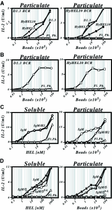
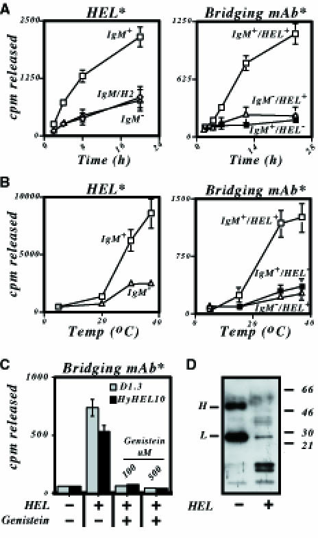
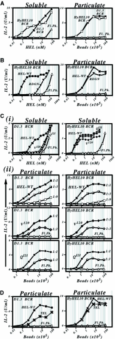
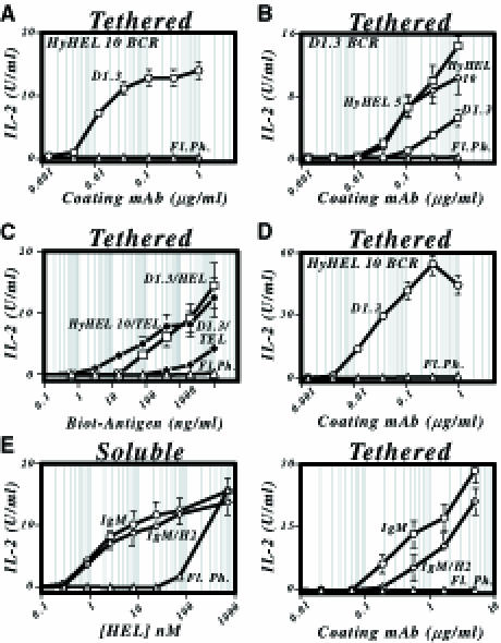
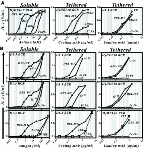
Binding of antigen to B-cell antigen receptor (BCR) leads to antigen internalization and presentation to T cells, a critical process in the initiation of the humoral immune response. However, antigen internalization has been demonstrated for soluble antigen, in vivo antigen is often encountered in insoluble form or tethered to a cell surface. Here, we show that not only can B cells internalize and present large particulate antigen (requiring a signalling-competent BCR to drive antigen uptake), but they can also extract antigen that is tethered tightly to a non-internalizable surface. The form in which the antigen is displayed affects the B cell's ability to discriminate antigen-BCR affinity. Thus, arraying an antigen on a particle or surface allows efficient presentation of low affinity antigens. However, the presentation efficiency of antigen arrayed on an internalizable particle plateaus at low affinity values. In contrast, extraction and presentation of antigen from a non-internalizable surface depends on antigen-BCR affinity over a wide affinity range. The results have implications for understanding both the initiation and affinity maturation of the immune response.


Figures

None
Fig. 1. Presentation of particulate antigen by B cells. (A) Presentation of HEL25–43/I-Ak by HEL-specific transfectants of the LK35.2 B–cell lymphoma that were incubated with HEL-coated beads was monitored by measuring IL-2 production from the co-cultured 2B6.3 T–cell hybridoma. The LK35.2 transfectants expressed either a D1.3 or a HyHEL10 BCR as indicated. HEL was bound onto streptavidin-coated beads via a biotinylated HyHEL10 (▪), HyHEL5 (•) or D1.3 (♦) mAb bridge. Untransfected LK35.2 B cells provided a control for presentation through fluid phase uptake (Fl. Ph., ▵) and give the same result as presentation by HEL-specific B–cell transfectants incubated in the absence of a bridging mAb. (B) Presentation of HEL108–116/I-Ad to the 1E5 T–cell hybridoma by A20[D1.3] (left-hand panel, ○) or A20[HyHEL10] (right-hand panel, □) B–cell transfectants that had been co-cultured with HEL covalently conjugated to tosyl-activated beads. Fluid phase presentation (▵). (C) Presentation of HEL1–18/I-Ek to the 2G7 T–cell hybridoma by transfectants of the LK35.2 B–cell lymphoma that express either the canonical D1.3 IgM HEL-specific BCR (○) or a D1.3 IgM/H2 chimera (⋄). Fluid phase presentation by untransfected LK35.2 cells (▵). Presentation was monitored using either soluble HEL (Soluble; left-hand panel) or biotinylated HEL bound to streptavidin-coated beads (Particulate; right-hand panel). (D) Presentation of HEL108–116/I-Ad to the 1E5 T–cell hybridoma by transfectants of the A20 B–cell lymphoma that express the canonical D1.3 IgM HEL-specific BCR (IgM; ▪), an IgM–β chimera (▪) or an IgM–β chimera carrying a Y→L mutation of the membrane-proximal cytoplasmic tyrosine (□). Fluid phase presentation by untransfected A20 cells (▵). Antigen was displayed in either soluble or particulate form as in (C).
None
Fig. 2. Degradation of particulate antigen by B cells. (A) Degradation of antigen or bridging mAb as a function of time. Transfectants of LK35.2 expressing either a canonical HEL-specific IgM BCR (IgM+; □), a HEL-specific IgM–H2 chimeric receptor (IgM/H2, ⋄) or no transfected BCR (IgM, ▵) were incubated at 37°C either with beads displaying [125I]HEL (left-hand panel) or with beads displaying non-radioactive HEL bound by an 125I-labelled bridging mAb (right-hand panel). Degradation of HEL or bridging mAb is presented as radioactivity released into the culture supernatant. In the right-hand panel, degradation of the bridging mAb was monitored in situations where it had (IgM+ HEL+; □ and ▵) or had not (IgM+ HEL; ▪) been pre-loaded with soluble HEL. Both the [125I]HEL in the right-hand panel and the 125I-labelled bridging mAb (HyHEL5) in the right-hand panel were covalently conjugated to beads. (B) Degradation of antigen or bridging antibody as a function of temperature. The experiment is as described in (A) except that the incubation was performed for 24 h at various temperatures. (C) Sensitivity of degradation to genistein. LK[HyHEL10] or LK[D1.3] transfectants were incubated for 6 h at 37°C with tosyl-activated beads containing 35S-labelled covalently conjugated HEL-specific bridging mAb (HyHEL5) which had or had not been pre-loaded with soluble HEL. The incubation was performed in the absence or presence of genistein and the results are presented as radioactivity released into the culture supernatant. (D) Degradation of bridging antibody detected by a SDS–PAGE on a 20% polyacrylamide gel. LK[HyHEL10] transfectants were incubated for 24 h at 37°C with a biotinylated HEL-specific bridging mAb (F10) immobilized on the surface of streptavidin-coated beads that had or had not been pre-loaded with soluble HEL. At the end of the incubation, the anti-HEL mAb was boiled off the beads, subjected to SDS–PAGE and detected by Western blotting with peroxidase-conjugated streptavidin.
None
Fig. 3. Dependence of the presentation of particulate antigen on antigen–BCR affinity. (A) Comparison of presentation of HEL108–116/I-Ad by LK35.2 B–cell transfectants carrying a HyHEL10 (□) or D1.3 (▪) BCR that have been co-cultured with HEL in either soluble (left panel) or particulate forms (right panel; covalently conjugated to tosyl-activated beads). (B) Comparison of presentation of wild-type HEL (▪) and of a mutant (HEL[R21, D101, G102, N103] designated RDGN, ▾) that shows reduced affinity for the HyHEL10 BCR by LK[HyHEL10] transfectants when the antigen is encountered either in soluble form (left-hand panel) or covalently conjugated to tosyl-activated beads (right-hand panel). Presentation of wild-type HEL by untransfected LK35.2 cells is shown as a control (▵). (C) Comparison of presentation of wild-type HEL (▪) and of two mutants displaying reduced affinity for the D1.3 BCR (HEL[V120], ♦; HEL[Q121], •) by LK[D1.3] transfectants. (i) The top pair of panels shows that presentation of these mutants through the D1.3 BCR (but not through the HyHEL10 BCR) is much reduced when they are encountered in soluble form. (ii) The lower panels show presentation of these mutant lysozymes through the D1.3 BCR (left-hand panels) as well as through the HyHEL10 BCR (control, right-hand panels) when encountered arrayed on a bead. The lysozymes were arrayed at various densities on streptavidin-coated beads by use of a biotinylated HEL-specific mAb bridge that was established by incubating 107 streptavidin-coated beads in 1 ml of PBS/BSA/Tween with biotinylated F10 mAb at concentrations of 5 (filled symbols), 1.67 (half-filled symbols) or 0.56 (open symbols) μg/ml prior to loading with saturating amounts of HEL. Presentation in (i) was monitored using 2B6 T cells, and in (ii) using 1E5 cells. HEL[Q121] gives a slightly reduced amplitude of IL-2 production from 1E5 T cells with both D1.3 and HyHEL10 transfectants, possibly reflecting the proximity of the Q121 mutation to the T–cell epitope recognized; this same reduction is not evident when the presentation of several other T–cell epitopes is monitored. (D) Presentation of HEL108–116/I-Ad by LK35.2 B–cell transfectants incubated with turkey egg lysozyme (TEL; ×) or HEL (▪) that have been covalently conjugated directly onto tosyl-activated beads. Presentation by LK[D1.3], left-hand panel; by LK[HyHEL10], right-hand panel. TEL, when encountered in solution, does not yield a level of presentation through the D1.3 BCR above that attributable to fluid phase uptake (see Figure 5D).
None
Fig. 4. Presentation by B cells of antigen immobilized on a plate. (A) Presentation of HEL108–116/I-Ad by LK[HyHEL10] B cells (○) that have been incubated on plates onto the surface of which HEL has been tethered through a D1.3 mAb bridge coated at various concentrations. Presentation by untransfected LK35.2 cells, ▵. (B) Presentation of HEL108–116/I-Ad by LK[D1.3] B cells that have been incubated on plates onto the surface of which HEL has been tethered through a HyHEL5 (□), HyHEL10 (⋄) or D1.3 (○) mAb bridge. (C) Presentation of HEL108–116/I-Ad by B–cell transfectants incubated on streptavidin-coated plates displaying biotinylated lysozyme. Presentation by LK[HyHEL10] transfectants incubated on plates displaying biotinylated HEL (•). Presentation by LK[D1.3] transfectants incubated on plates displaying biotinylated HEL (□) or TEL (♦). Plates were coated with streptavidin (20 μg/ml) in PBS and, after washing and blocking, incubated with biotinylated HEL/TEL at the indicated concentration. (D) Presentation of HEL108–116/I-Ad by LK[HyHEL10] B cells (○) that have been incubated on Reacti-Bind™ plates which display HEL tethered by covalently linked D1.3 mAb. (E) Presentation of HEL1–18/I-Ek by HEL-specific transfectants of LK35.2 that have been incubated with either soluble HEL (left-hand panel) or HEL tethered onto HyHEL5-coated plastic plates (right-hand panel). Presentation by LK[D1.3 canonical BCR], ○; by LK[D1.3 IgM–H2 chimeric BCR], ⋄; untransfected LK35.2 cells, ▵.
None
Fig. 5. Dependence of the presentation of plate-immobilized antigen on antigen–BCR affinity. (A) Presentation of lysozyme mutants showing a diminished affinity for HyHEL10. The left-hand panel shows presentation by LK[HyHEL10] B–cell transfectants of the antigens when encountered in solution; the middle and right-hand panels show presentation by LK[HyHEL10] or LK[D1.3] transfectants of the same antigens when displayed immobilized on a plate tethered by an anti-HEL mAb (D1.3 tether, middle panel; HyHEL5 tether, right-hand panel). Wild-type HEL, ▪; HEL[R21, D101], ○× (RD); HEL[R21, D101, G102, N103], ▾ (RDGN); untransfected LK35.2 control, ▵. (B) Presentation of lysozyme variants showing a diminished affinity for D1.3. The left-hand panel shows presentation by LK[D1.3] B–cell transfectants of the antigens when encountered in solution; the middle and right-hand panels show presentation by LK[D1.3] or LK[HyHEL10] B–cell transfectants of the same antigens when displayed tethered to the plate via HyHEL5 mAb. Wild-type HEL, ▪; HEL[V120], ♦; HEL[Q121], •; TEL, ×; untransfected LK35.2 control, ▵. Presentation was monitored using 2G7 T cells, except in the experiment investigating presentation of soluble HEL[V120], where 2B6 was used.

Similar articles

Cited by

References

    1. Adorini L., Guery, J.C., Fuchs, S., Ortiz, N.V., Hammerling, G.J. and Momburg, F. (1993) Processing of endogenously synthesized hen egg-white lysozyme retained in the endoplasmic reticulum or in secretory form gives rise to a similar but not identical set of epitopes recognized by class II-restricted T cells. J. Immunol., 151, 3576–3586. - PubMed
    1. Aluvihare V.R., Khamlichi, A.A., Williams, G.T., Adorini, L. and Neuberger, M.S. (1997) Acceleration of intracellular targeting of antigen by the B–cell antigen receptor: importance depends on the nature of the antigen–antibody interaction. EMBO J., 16, 3553–3562. - PMC - PubMed
    1. Batista F.D and Neuberger, M.S. (1998) Affinity dependence of the B cell response to antigen: a threshold, a ceiling and the importance of off-rate. Immunity, 8, 751–759. - PubMed
    1. Daeron M. (1997) Fc receptor biology. Annu. Rev. Immunol., 15, 203–234. - PubMed
    1. Davies D.R. and Padlan, E.A. (1990) Antibody–antigen complexes. Annu. Rev. Biochem., 59, 439–473. - PubMed

Publication types