Comparative Study
. 2000 Feb 15;19(4):542-9.
doi: 10.1093/emboj/19.4.542.

YidC, the Escherichia coli homologue of mitochondrial Oxa1p, is a component of the Sec translocase

Affiliations
Comparative Study

YidC, the Escherichia coli homologue of mitochondrial Oxa1p, is a component of the Sec translocase

P A Scotti et al. EMBO J. .

Abstract

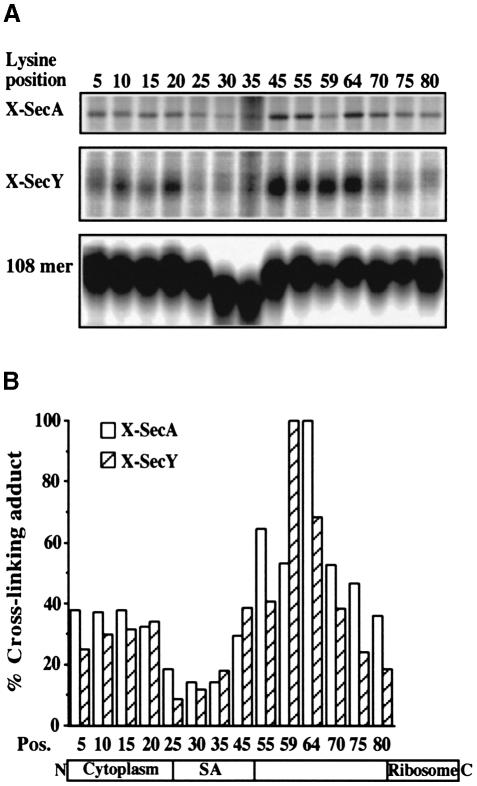
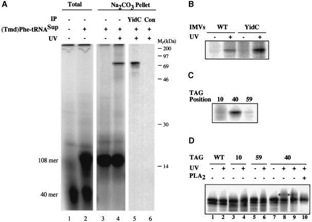
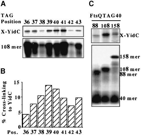
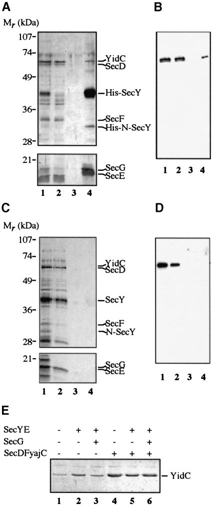
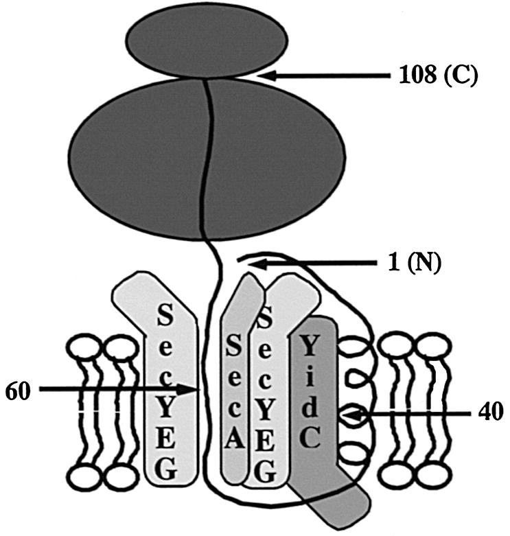
In Escherichia coli, both secretory and inner membrane proteins initially are targeted to the core SecYEG inner membrane translocase. Previous work has also identified the peripherally associated SecA protein as well as the SecD, SecF and YajC inner membrane proteins as components of the translocase. Here, we use a cross-linking approach to show that hydrophilic portions of a co-translationally targeted inner membrane protein (FtsQ) are close to SecA and SecY, suggesting that insertion takes place at the SecA/Y interface. The hydrophobic FtsQ signal anchor sequence contacts both lipids and a novel 60 kDa translocase-associated component that we identify as YidC. YidC is homologous to Saccharomyces cerevisiae Oxa1p, which has been shown to function in a novel export pathway at the mitochondrial inner membrane. We propose that YidC is involved in the insertion of hydrophobic sequences into the lipid bilayer after initial recognition by the SecAYEG translocase.


Figures

None
Fig. 1. Scanning cross-linking of single lysine 108FtsQ mutants to SecA and SecY. (A) Mutant 108FtsQ was synthesized in the presence of IMVs. The samples were treated with the bifunctional cross-linker DSS and extracted with sodium carbonate. Panels corresponding to SecA and SecY cross-linking adducts (X-SecA and X-SecY) and nascent chains (108mer) are shown. The latter panel is derived from a separate 15% gel to allow a better resolution and quantification of small polypeptides (see Materials and methods). (B) SecA and SecY cross-linking adducts from (A) were quantified by phosphoimaging and each signal was corrected for the respective translation efficiency. Highest values for cross-linking efficiency were taken as 100%. Average values of three independent experiments are shown. The positions of the lysine residues in the mutant 108FtsQ as well as their location in the different domains of 108FtsQ are indicated.
None
Fig. 2. The SA sequence of targeted 108FtsQ interacts with both YidC and lipids. (A) In vitro translation of 108FtsQTAG40 was carried out in the presence of IMVs and in the absence or presence of (Tmd)Phe-tRNASup as indicated. Aliquots were TCA precipitated (Total). Upon translation in the presence of (Tmd)Phe-tRNASup, samples were UV irradiated or kept in the dark for 10 min at 4°C and extracted with sodium carbonate as indicated. UV-irradiated samples were immunoprecipitated using antiserum raised against YidC or control pre-immune serum as indicated. (B) IMVs from a strain that overproduces YidC were compared with wild-type IMVs in the photocross-linking reaction described in (A). YidC adducts in the sodium carbonate pellet are shown. (C) 108FtsQTAG10 and 59 were compared with 108FtsQTAG40 in the photocross-linking reaction described in (A). The amounts of suppressed 108FtsQ produced during translation were equalized to enable direct visual comparison of cross-linking efficiencies. YidC cross-linking adducts in the sodium carbonate pellet are shown. (D) 108FtsQ nascent chains were produced and cross-linked as described in (A) with (Tmd)Phe incorporated at positions 10, 40 and 59 as indicated. As a control, wild-type 108FtsQ (WT) was produced under the same translation conditions. Prior to sodium carbonate extraction, samples were UV irradiated or kept in the dark as indicated. As a control, 108FtsQTAG40 nascent chains were treated with bee venom phospholipase (PLA2; lane 10) or mock treated in incubation buffer (lane 9). Lipid cross-linking adducts are indicated by an asterisk.
None
Fig. 3. Scanning photo cross-linking of targeted FtsQ mutants to YidC. (A) TAG codons at positions 36–43 in the SA sequence of 108FtsQ were suppressed as described in the legend to Figure 2. Half of each sample was kept in the dark and extracted with sodium carbonate to determine the amount of suppressed 108FtsQ present in each membrane fraction (108mer panel). The other half was UV irradiated for 10 min prior to extraction. YidC cross-linking adducts in sodium carbonate-extracted membranes are shown (X-YidC panel). (B) YidC cross-linking adducts from (A) were quantified and expressed relative to the amount of suppressed 108FtsQ present prior to cross-linking in the sodium carbonate-extracted membranes. The average values of three independent experiments are shown. (C) FtsQ constructs of different length with a TAG codon at position 40 were suppressed as described in the legend to Figure 2. After translation, aliquots were taken and either directly TCA precipitated to determine suppression and translation efficiencies (lower panel) or extracted with sodium carbonate to determine the integration efficiency of each construct into the membrane (not shown). The rest of the translation reactions were UV irradiated for 10 min (the same amount of nascent chains was present in each sample prior to cross-linking). YidC cross-linking adducts in sodium carbonate-extracted membrane pellets are shown (X-YidC panel).
None
Fig. 4. YidC is associated with the SecYEG complex. YajCSechisYEGDF (A and B) and YajCSecYEGDF (C and D) IMVs solubilized with DDM and partially purified by DEAE anion exchange chromatography (lane 1) loaded on an Ni+–NTA column (lane 2, flow-through), washed (lane 3, wash) and eluted with imidazole (lane 4, elution). Samples were analysed by SDS–PAGE and silver staining (A and C). YidC was detected using Western blotting with the YidC polyclonal antiserum (B and D). The positions of the various proteins are indicated. His-N-SecY is a proteolytic N–terminal fragment of SecY. (E) IMVs derived from the wild-type cells (lane 1) and cells overproducing SecYE (lane 2), SecYEG (lane 3), YajCSecDF (lane 4), YajCSecYEDF (lane 5) or YajCSecYEGDF (lane 6) were isolated. Samples were loaded on SDS–PAGE gels and the levels of YidC were detected with the YidC polyclonal antiserum after Western blotting.
None
Fig. 5. Model for the molecular environment of membrane-inserted nascent 108FtsQ.

References

    1. Brunner J. (1996) Use of photocrosslinkers in cell biology. Trends Cell Biol., 6, 154–157. - PubMed
    1. Cagnon C., Valverde, V. and Masson, J.-M. (1991) A new family of sugar-inducible expression vectors for Escherichia coli.Protein Eng., 4, 843–847. - PubMed
    1. Carson M.J., Barondess, J. and Beckwith, J. (1991) The FtsQ protein of Escherichia coli: membrane topology, abundance and cell division phenotypes due to overproduction and insertion mutations. J. Bacteriol., 173, 2187–2195. - PMC - PubMed
    1. De Gier J.W.L., Mansournia, P., Valent, Q.A., Phillips, G.J., Luirink, J. and von Heijne, G. (1996) Assembly of a cytoplasmic membrane protein in Escherichia coli is dependent on the signal recognition particle. FEBS Lett., 399, 307–309. - PubMed
    1. De Vrije T., Tommassen, J. and de Kruijff, B. (1987) Optimal posttranslational translocation of the precursor of PhoE protein across Escherichia coli membrane vesicles requires both ATP and the proton motive force. Biochim. Biophys. Acta, 900, 63–72. - PubMed

Publication types

MeSH terms