. 2000 Feb;105(3):253-60.
doi: 10.1172/JCI7630.

NF-kappaB determines localization and features of cell death in epidermis

Affiliations

NF-kappaB determines localization and features of cell death in epidermis

C S Seitz et al. J Clin Invest. 2000 Feb.

Abstract

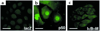
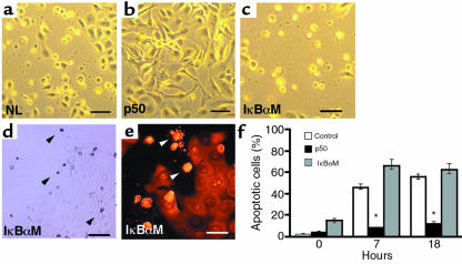
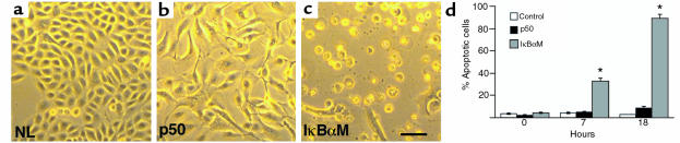
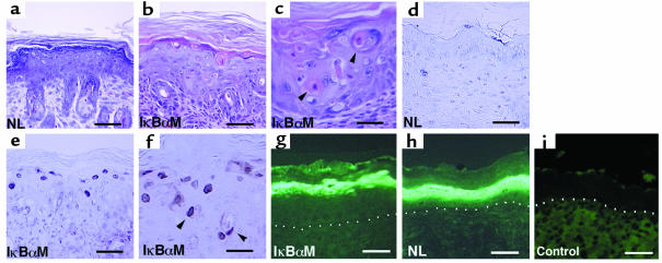
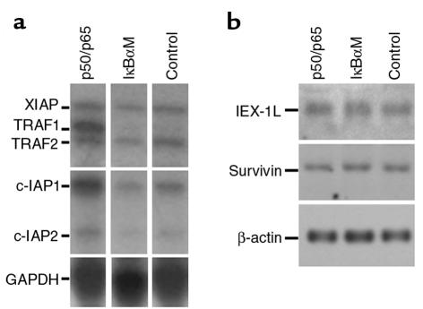
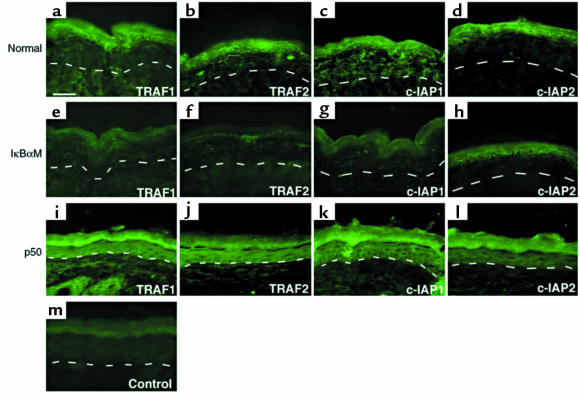
Specialized forms of physiologic cell death lacking certain characteristic morphologic features of apoptosis occur in terminally differentiating tissues, such as in the outer cell layers of epidermis. In these cell layers, NF-kappaB translocates from the cytoplasm to the nucleus and induces target gene expression. In light of its potent role in regulating apoptotic cell death in other tissues, NF-kappaB activation in these cells suggests that this transcription factor regulates cell death during terminal differentiation. Here, we show that NF-kappaB protects normal epithelial cells from apoptosis induced by both TNFalpha and Fas, whereas NF-kappaB blockade enhances susceptibility to death via both pathways. Expression of IkappaBalphaM under control of keratin promoter in transgenic mice caused a blockade of NF-kappaB function in the epidermis and provoked premature spontaneous cell death with apoptotic features. In normal tissue, expression of the known NF-kappaB-regulated antiapoptotic factors, TRAF1, TRAF2, c-IAP1, and c-IAP2, is most pronounced in outer epidermis. In transgenic mice, NF-kappaB blockade suppressed this expression, whereas NF-kappaB activation augmented it, consistent with regulation of cell death by these NF-kappaB effector proteins. These data identify a new role for NF-kappaB in preventing premature apoptosis in cells committed to undergoing physiologic cell death and indicate that, in stratified epithelium, such cell death normally proceeds via a distinct pathway that is resistant to NF-kappaB and its antiapoptotic target effector genes.


Figures

Figure 1
Figure 1
Generation of epithelial cells with altered NF-κB function. Normal keratinocytes were transduced with retroviral expression vector for (a) lacZ normal control, (b) constitutively active p50, and (c) the trans-dominant IκBαM super-repressor, then subjected to immunofluorescence staining with antibody to p50 (bars = 5 μm). Note marked nuclear expression in p50-transduced cells and blockade of nuclear-localized p50 in IκBαM.
Figure 2
Figure 2
NF-κB subunit protects epithelial cells against Fas-induced apoptosis in vitro. Normal human keratinocytes were cotransduced with a retroviral vector of the wild-type human Fas protein along with vectors for p50, IκBαM, and lacZ normal control (NL). One day after double transduction, Fas signal transduction was triggered by the anti–Fas antibody CH-11 (1 μg/mL) and cellular morphology was analyzed by phase-contrast microscopy (bars = 10 μm). (ac) Morphology of (a) normal control, (b) p50[+], and (c) IκBαM[+] cells 7 hours after Fas activation. Note the rounded, shrunken, and detached morphology of IκBαM[+] and normal controls and the lack of such changes in p50[+] cells. Representative (d) TUNEL assay stains and (e) propidium iodide nuclear stains demonstrate apoptotic changes in cells with altered morphology; note positive nuclei in TUNEL staining as well as nuclear condensation and collapse evident in propidium iodide stained cells (arrows). (f) Quantitation of NF-κB effects on Fas-induced epithelial apoptosis in vitro. The percentage of apoptotic cells was determined by cell morphology, TUNEL, and nuclear stains. Time points analyzed were 0, 7, and 18 hours after Fas cross-linking with CH-11. Three independent transductions were analyzed at each time point; data shown are based on morphologic changes and are presented as ± SD between these 3 independent experiments. *P < 0.01 difference of p50 from IκBαM and normal controls.
Figure 3
Figure 3
NF-κB blockade renders normal epithelial cells susceptible to TNFα-triggered apoptosis. Normal keratinocytes were transduced with retroviral expression vectors for p50, IκBαM, and lacZ normal control (NL), then treated 24 hours later with TNFα. (a) Normal control and (b) p50[+] cells; note failure to demonstrate cellular rounding, shrinkage, and detachment characteristic of apoptosis in contrast to (c) IκBαM[+] cells (bar = 10 μm). (d) Quantitation of NF-κB effects on TNFα-triggered epithelial apoptosis in vitro. The percentage of apoptotic cells was determined by cell morphology, TUNEL, and nuclear stains. Time points analyzed were 0, 7, and 18 hours after addition of TNFα. Three independent transductions were analyzed at each time point; data shown are based on morphologic changes and are presented as ± SD between these 3 independent experiments. *P < 0.01 difference of IκBαM from p50 and controls.
Figure 4
Figure 4
Epidermal apoptosis with blockade of NF-κB function in vivo. (ac) Histology of skin in vivo. (a) Age- and site-matched control versus (b, c) IκBαM[+] mice transgenic for loss of epidermal NF-κB function. Note the presence of apoptotic cell morphologic changes extending down to the lower spinous layers (arrows) and the complete absence of such changes in the control. (df) TUNEL assay in vivo. (d) Age- and site-matched control versus (e, f) mice transgenic for loss of epidermal NF-κB function. Note the presence of TUNEL-positive cells in the granular layer extending down into the lower spinous layers in IκBαM[+] mice. (a, b, d, e) Bars = 50 μm, (c, f) bars = 25 μm. Expression of the terminal differentiation marker loricrin is not inhibited by NF-κB blockade in vivo. Skin from (g) keratin promoter–driven IκBαM[+] transgenic mice (NL) (h) and littermate control were immunostained using antibody specific for loricrin. (i) Anti-mouse secondary antibody alone controls for background immunofluorescence. The dermal-epidermal boundary is highlighted by white dots. (g, h) Bars = 40 μm.
Figure 5
Figure 5
NF-κB effects on expression of antiapoptotic genes. (a) Ribonuclease protection. Normal human keratinocytes were transduced with retroviral expression vectors for p50 and p65 (p50/p65), IκBαM, and lacZ normal control (NL). RNA extracted 12 hours later and subjected to multi-gene ribonuclease protection assay. (b) Northern analysis. Survivin and IEX-1L mRNA expression was determined in human keratinocytes expressing p50 and p65 (p50/p65), IκBαM, and lacZ normal control.
Figure 6
Figure 6
NF-κB effects on expression of antiapoptotic genes in mice transgenic for augmentation or loss of epidermal NF-κB function. (ad) Immunofluorescence analysis of TRAF1, TRAF2, c-IAP1, and c-IAP2 in normal littermate control. (eh) Immunofluorescence analysis in IκBαM[+] transgenic mice. (il) Immunofluorescence analysis in mice transgenic for constitutively active p50 NF-κB subunit; note relative hypoplasia of p50[+] epidermis. (m) Anti-rabbit secondary antibody alone control. Note decrease in detection of TRAF1, TRAF2, c-IAP1, and, to a lesser extent, c-IAP2 in IκBαM[+] epidermis, especially in the outer epithelial layers, compared with control. Note also the marked increase in TRAF1, TRAF2, c-IAP1, and, to a lesser extent, c-IAP2 expression, throughout epidermis of p50[+] skin, including cells all the way to the basal layer. The dashed line represents the epidermal basement membrane zone; all layers of epidermal morphology are highlighted in p50[+] skin. All micrographs are at the same magnification (bar = 50 μM, shown in a).

References

    1. Tamada Y, et al. Identification of programmed cell death in normal human skin tissues by using specific labelling of fragmented DNA. Br J Dermatol. 1994;131:521–524. - PubMed
    1. Polakowska R, Piacentini M, Bartlett R, Goldsmith L, Haake AR. Apoptosis in human skin development: morphogenesis, periderm, and stem cells. Dev Dyn. 1994;199:176–188. - PubMed
    1. Vaux DL, Korsmeyer SJ. Cell death in development. Cell. 1999;96:245–254. - PubMed
    1. Haake AR, Polakowska RR. Cell death by apoptosis in epidermal biology. J Invest Dermatol. 1993;101:107–112. - PubMed
    1. Hill LL, et al. Fas ligand: a sensor for DNA damage critical in skin cancer etiology. Science. 1999;285:898–900. - PubMed

Publication types

MeSH terms