Review
. 2000 Feb;105(3):261-70.
doi: 10.1172/JCI8856.

The basic helix-loop-helix transcription factor, dHAND, is required for vascular development

Affiliations
Review

The basic helix-loop-helix transcription factor, dHAND, is required for vascular development

H Yamagishi et al. J Clin Invest. 2000 Feb.

Abstract

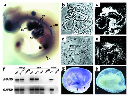
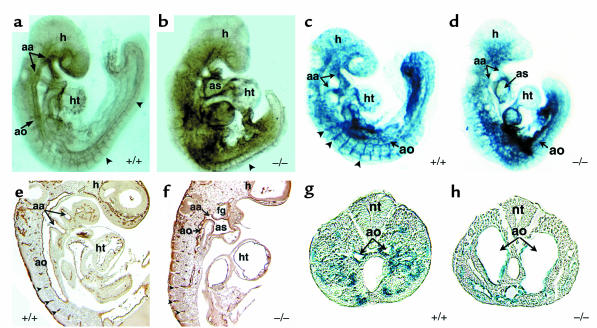
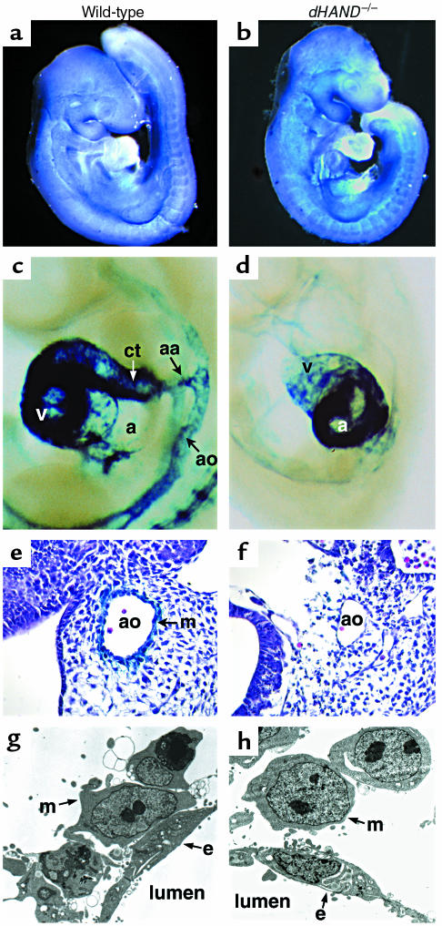
Reciprocal interactions between vascular endothelial cells and vascular mesenchymal cells are essential for angiogenesis. Here we show that the basic helix-loop-helix transcription factor, dHAND/Hand2, is expressed in the developing vascular mesenchyme and its derivative, vascular smooth muscle cells (VSMCs). Targeted deletion of the dHAND gene in mice revealed severe defects of embryonic and yolk sac vascular development by embryonic day 9.5. Vascular endothelial cells expressed most markers of differentiation. Vascular mesenchymal cells migrated appropriately but failed to make contact with vascular endothelial cells and did not differentiate into VSMCs. In a screen for genes whose expression was dependent upon dHAND (using subtractive hybridization comparing wild-type and dHAND-null hearts), the VEGF(165) receptor, neuropilin-1, was found to be downregulated in dHAND mutants. These results suggest that dHAND is required for vascular development and regulates angiogenesis, possibly through a VEGF signaling pathway.


Figures

Figure 1
Figure 1
Expression of dHAND in the developing vasculature. Whole-mount in situ hybridization revealed dHAND expression in the aortic arch arteries (aa) and aorta (ao) of an E10.0 embryo (a). Expression in the pharyngeal arches (pa), heart (ht), and limb bud (lb) was also seen. Sagittal section of an E9.5 embryo (b and c) showed expression in the aorta and aortic arch arteries. Sagittal section of an E10.5 embryo (d and e) demonstrated dHAND expression in the mesenchyme (m) of the third and fourth aortic arch arteries (arrowheads) and in the pharyngeal arch (pa) mesenchyme. b and d represent phase images of c and e, respectively. Expression of dHAND was also observed in the vascular smooth muscle cell line, A10, and in the adult heart and aorta by RT-PCR (f). RNA integrity was assayed by amplification of GAPDH. Yolk sac expression of dHAND was observed by RT-PCR in f and whole-mount in situ hybridization (arrowheads indicate vessels) (g). (h) Sense control.
Figure 2
Figure 2
Endothelial development in dHAND mutants. Whole-mount immunochemistry revealed that endothelial cells expressed PECAM-1 protein appropriately in wild-type (a) and mutant (b) E9.5 embryos, but displayed a disorganized pattern in dHAND mutants (b). The rostral portion of dHAND-null embryos was more severely affected than the caudal region, where the aorta (ao) and somitic arteries (arrowheads) were visible. Sagittal section of PECAM-1 antibody–stained wild-type (e) and mutant (f) embryos revealed disorganization of the dorsal aorta of dHAND-null embryos, where the aortic lumen was evident. Note patency of the aortic arch artery (aa) in the mutant (f). β-galactosidase activity in wild type (c) and mutant (d) embryos harboring lacZ under control of the Tie2 promoter revealed disorganization of lacZ expression in dHAND-null embryos compared with wild-type embryos. Transverse sections of wild-type (g) and mutant (h) embryos in the caudal region demonstrate the dilated nature of caudal vessels of dHAND-null embryos. ht, heart; as, aortic sac; h, head; nt, neural tube; fg, foregut.
Figure 3
Figure 3
Normal endothelial cell differentiation in dHAND-null embryos. Semiquantitative RT-PCR analysis of wild-type (+/+) and dHAND-null (–/–) embryos using primers specific to the endothelial cell markers Flk-1, Tie1, Tie2, Flt-1, and VEGF revealed equal levels of expression suggesting normal differentiation and quantity of endothelial cells in dHAND mutants. Similarly, the mesenchymal marker, Ang-1, was unaffected in dHAND mutants, as were the transcription factors MEF2C and ARNT. GAPDH expression indicates equal RNA loading.
Figure 4
Figure 4
Yolk sac vascular defects in dHAND-null embryos. Unlike wild-type yolk sacs at E9.5 (a), dHAND-null yolk sacs at E9.5 display absence of visible vessels (b). Expression of lacZ under control of the Tie2 promoter in wild-type (c) and mutant (d) yolk sacs revealed a honeycomb-like vascular plexus in dHAND-mutants rather than the remodeled vessels present in the wild-type (e) and (f) represent histologic sections of c and d, respectively. Arrowheads indicate vessels; arrows indicate endothelial cells.
Figure 5
Figure 5
Failure of vascular smooth muscle differentiation in dHAND mutants. Mesenchymal cells in wild-type (a) and dHAND mutants (b) expressed the mesenchymal marker, COUPTFII, throughout the embryo. Expression of lacZ under the control of the SM-22α promoter was seen in the aortic arch arteries (aa), aorta (ao), conotruncus (ct), and ventricle (v) of the heart of an E9.5 transgenic embryo (c). In the dHAND-null background, the SM-22-lacZ transgene expressed in the atrium (a) of the heart, but not in the vasculature (d) at E9.5. Embryos are shown in lateral views focusing on the cardiac and vascular areas. Histologic analysis of wild-type (e) and mutant (f) embryos revealed absence of lacZ expression in the cells surrounding the aorta in dHAND-null embryos. Electron microscopy of transverse sections through the rostral part of E9.5 wild-type (g) and dHAND-null (h) embryos revealed vascular mesenchymal (m) cells surrounding endothelial (e) cells. Wild-type mesenchymal cells developed cytoplasmic processes to contact endothelial cells, whereas dHAND-null mesenchymal cells remained rounded without contacting endothelial cells.
Figure 6
Figure 6
Neuropilin-1 is downregulated in dHAND mutants. Neuropilin-1 was isolated as a dHAND-dependent gene by subtractive hybridization between dHAND-mutant and wild-type hearts (a). Quantitative RT-PCR of neuropilin-1 in wild-type (+/+) and dHAND-null (–/–) hearts confirmed downregulation of neuropilin-1 in dHAND-mutant heart and yolk sac RNA (b). Cycles of PCR amplification are shown. RNA loading was controlled by assaying GAPDH expression. Whole-mount in situ hybridization demonstrated neuropilin-1 expression in the heart (ht) and aorta (arrowheads) of an E9.0 embryo in right lateral view (c). At E9.5 (d), neuropilin-1 expression, viewed from the left lateral side, was seen in the aorta and aortic arch arteries (arrowheads), heart, pharyngeal arch (pa), limb bud (lb), and septum transversum (st). In dHAND mutants, neuropilin-1 was downregulated in the rostral aorta, pharyngeal arches, and heart (e). Expression in the caudal aorta (arrowhead), limb bud, and septum transversum was intact. Transverse sections through the rostral part of embryos in d and e revealed neuropilin-1 expression in the endothelial (e) and mesenchymal (m) cells of the wild-type aorta (f), but no expression of neuropilin-1 in the dHAND-null aorta (g). h, head.

References

    1. Folkman J, D’Amore PA. Blood vessel formation: what is its molecular basis? Cell. 1996;87:1153–1155. - PubMed
    1. Risau W, Flamme I. Vasculogenesis. Annu Rev Cell Dev Biol. 1995;11:73–91. - PubMed
    1. Risau W. Mechanisms of angiogenesis. Nature. 1997;386:671–674. - PubMed
    1. Hanahan D. Signaling vascular morphogenesis and maintenance. Science. 1997;277:48–50. - PubMed
    1. Dumont DJ, et al. Dominant-negative and targeted null mutations in the endothelial receptor tyrosine kinase, tek, reveal a critical in vasculogenesis of the embryo. Genes Dev. 1994;8:1897–1909. - PubMed

Publication types

MeSH terms