. 2000 Feb;105(3):301-10.
doi: 10.1172/JCI7358.

Differential immune responses to alpha-gal epitopes on xenografts and allografts: implications for accommodation in xenotransplantation

Affiliations

Differential immune responses to alpha-gal epitopes on xenografts and allografts: implications for accommodation in xenotransplantation

M Tanemura et al. J Clin Invest. 2000 Feb.

Abstract

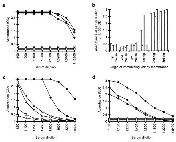
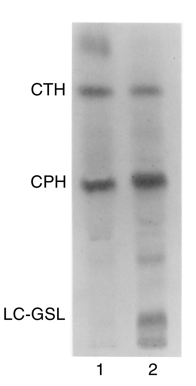
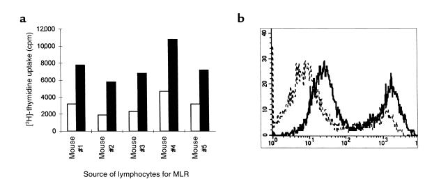
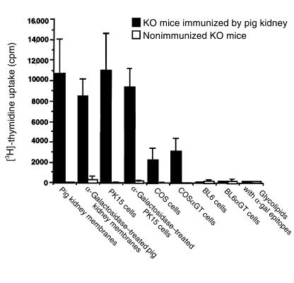
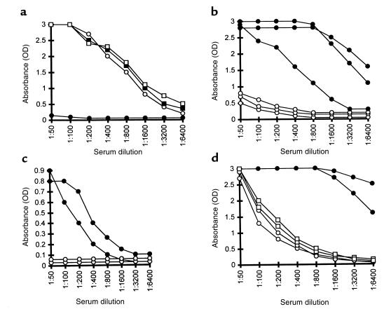
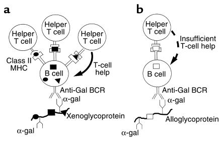
Xenograft recipients produce large amounts of high-affinity anti-Gal IgG in response to Galalpha1-3Galbeta1- 4GlcNAc-R (alpha-gal) epitopes on the graft. In contrast, ABO-mismatched allograft recipients undergo "accommodation," a state of very weak immune response to ABO antigens. These differences in anti-carbohydrate immune response were studied in alpha1,3galactosyltransferase knock-out mice. Pig kidney membranes administered to these mice elicited extensive production of anti-Gal IgG, whereas allogeneic kidney membranes expressing alpha-gal epitopes elicited only a weak anti-Gal IgM response. Anti-Gal IgG response to xenograft membranes depended on helper T cell activation and was inhibited by anti-CD40L antibody. These T cells were activated by xenopeptides and not by alpha-gal epitopes. Moreover, allogeneic cell membranes manipulated to express xenoproteins also induced anti-Gal IgG response. Xenoglycoproteins with alpha-gal epitopes are processed by anti-Gal B cells. Xenopeptides presented by these cells activate a large repertoire of helper T cells required for the differentiation of anti-Gal B cells into cells secreting anti-Gal IgG. Alloglycoproteins with alpha- gal epitopes have very few immunogenic peptides and fail to activate helper T cells. Similarly, ineffective helper T-cell activation prevents a strong immune response to blood group antigens in ABO-mismatched allograft recipients, thus enabling the development of accommodation.


Figures

Figure 1
Figure 1
(a) Anti-Gal IgG production in α1,3GT KO mice immunized 3 times with kidney membranes from pig (filled circles) or C3H mouse (open circles). Data represent anti-Gal activity in 3 characteristic mice of 8 in each group with similar results. (b) Anti-Gal IgG activity in 5 mice immunized first 3 times with WT mouse kidney membranes followed by 3 immunizations with pig kidney membranes. Data represent absorbance 2 weeks after each immunization at serum dilution of 1:100. (c) Anti-Gal IgM response in nonimmunized α1,3GT KO mice (open squares), mice immunized 3 times with pig kidney membranes (filled circles), or with WT mouse kidney membranes (open circles). Data from 2 representative mice from groups of 5 animals per group. (d) Anti-Gal IgM (filled circles) and IgG (open circles) response in mice immunized 3 times with glycolipids containing α-gal epitopes in amounts equivalent to the amount of this epitope in 100 mg pig kidney membranes. Data are from 3 representative mice of 6 per group with similar results.
Figure 2
Figure 2
Thin-layer chromatography separation of rabbit RBC glycolipids extracted in the organic (lane 1) and aqueous phase (lane 2) after Folch partition. Glycolipids were stained with orcinol. Note that the organic phase contains primarily ceramide trihexoside (CTH) with the structure Galα1-4Galβ1-4Glc-Cer, whereas the aqueous phase contains mostly CPH with the structure Galα1-3Galβ1- 4GlcNAcβ1-3Galβ1-4Glc-Cer, and long-chain glycosphingolipids (LC-GSL), all of which have α-gal epitopes (15, 16).
Figure 3
Figure 3
Allogeneic response of α1,3GT KO mice to C3H mice. (a) Stimulation of lymphocytes from 5 α1,3GT KO mice in a MLR with irradiated C3H mouse lymphocytes as stimulatory cells (closed columns) or in the absence of stimulatory cells (open columns). (b) Production of alloantibodies in α1,3GT KO mice immunized with C3H kidney membranes; FACS analysis of C3H spleen lymphocytes incubated with sera from nonimmunized α1,3GT KO mice (broken line), or sera from immunized mice (solid line). The left peak in each histogram represents T cells, whereas the right peak represents B cells. Data are from 1 of 5 mice with similar results.
Figure 4
Figure 4
Two-color FACS analysis of B cells with anti-Gal BCR in α1,3GT KO mice. B cells were identified by the staining with PE-conjugated anti-mouse Ig, whereas cells with anti-Gal BCR were identified by the binding of biotinylated α-gal BSA, followed by avidin-FITC. The stained lymphocytes were from: (a) nonimmunized mice; (b) mice immunized with pig kidney membranes; (c) cells from mice immunized as in b and tested for binding of biotinylated N-acetyllactosamine (Galβ1-4GlcNAc-R)-BSA as specificity control; (d) mice immunized with WT mouse kidney membranes; (e) mice immunized with glycolipids from rabbit RBC. Proportion of double-positive stained cells is indicated. Data are from a representative mouse of 5 in each group.
Figure 5
Figure 5
Effect of α-gal epitope expression on in vitro proliferation of spleen lymphocytes from nonimmunized α1,3GT KO mice (open columns) or from α1,3GT KO mice immunized 3 times with pig kidney membranes (closed columns). Pig kidney membranes or pig PK15 cells were treated with α-galactosidase for the removal of α-gal epitopes. COS α GT and BL6 α GT cells are transfected with mouse α1,3GT cDNA and express α-gal epitopes. Glycolipids extracted from rabbit RBC were assayed at concentrations that corresponded to α-gal epitope expression by 100 μg/mL of pig kidney membranes. Data are mean ± SE of from 5 mice in each group.
Figure 6
Figure 6
Anti-Gal IgG response in α1,3GT KO mice is dependent on helper T cells that are activated by xenopeptides. (a) Effect of anti-CD40L antibody on anti-Gal production in mice immunized twice with pig kidney membranes: IgG (filled circles) and IgM (open circles) indicate response in mice treated with anti-CD40L antibody or with control hamster IgG (filled squares, open squares), respectively. Data are from a representative mouse of 5 in each group. (b) Response in mice immunized with human/mouse hybridoma (filled circles) or with mouse/mouse hybridoma (open circles). Data represent anti-Gal activity in 3 mice of 7 with similar results in each group. (c) Response in mice that were preimmunized with KLH, then immunized with KLH-coupled C3H mouse kidney membranes (filled circles) or with C3H kidney membranes (open circles). Data are from 2 representative mice of 4 in each group. (d) Response in mice preimmunized twice with pig kidney membranes and, after 2 months, immunized twice with allogeneic mouse kidney membranes (open circles) or with pig kidney membranes (filled circles). Antibody activity before second set of immunizations is represented by open squares. Data are from 2 representative mice of 5 in each group.
Figure 7
Figure 7
Destruction of cells expressing or lacking α-gal epitopes by the ADCC mechanism with mouse splenocytes as effector cells and with mouse anti-Gal (4 μg/mL). Target cells studied are: BL6α GT melanoma cells expressing α-gal epitopes (filled triangles); BL6 cells lacking α-gal epitopes (open triangles); COSα GT monkey cells expressing α-gal epitope (filled squares); COS cells lacking α-gal epitopes (open squares); pig PK15 cells (filled diamonds). Data are representative from 1 of 5 mice with similar results.
Figure 8
Figure 8
Proposed mechanism for the differential immune response to α-gal epitopes on xenografts and allografts. (a) Activation of anti-Gal B cells by xenoglycoproteins expressing α-gal epitopes and containing multiple immunogenic xenopeptides (filled circles, filled squares, filled triangles). The xenoglycoproteins are internalized by the anti-Gal B cell, subsequent to interaction of α-gal epitopes with anti-Gal BCR. The processed xenopeptides are presented in association with class II MHC molecules and effectively activate many helper T cells with the corresponding TCR specificities. These activated T cells provide the help required by the B cell to complete its activation and undergo proliferation, isotype switch, and affinity maturation for the ultimate production of high-affinity anti-Gal IgG. (b) The processing and presentation of relatively few immunogenic allopeptides (open squares) on allograft glycoproteins with α-gal epitopes results in insufficient helper T-cell activation. Thus, the anti-Gal B cell fails to complete its activation and to differentiate into a plasma cell producing IgG molecules.

References

    1. Galili U. Interaction of the natural anti-Gal antibody with α-galactosyl epitopes: a major obstacle for xenotransplantation in humans. Immunol Today. 1993;14:480–482. - PubMed
    1. Good AH, et al. Identification of carbohydrate structures that bind human antiporcine antibodies: implications for discordant xenografting in humans. Transplant Proc. 1992;24:559–562. - PubMed
    1. Sandrin MS, Vaughan HA, Dabkowski PL, McKenzie IFC. Anti-pig IgM antibodies in human serum react predominantly with Gal(α1-3)Gal epitopes. Proc Natl Acad Sci USA. 1993;90:11391–11395. - PMC - PubMed
    1. Cozzi, E., et al. 1997. Effect of transgenic expression of human decay-accelerating factor on the inhibition of hyper acute rejection of pig organs. In Xenotransplantation. 2nd edition. D.K.C. Cooper, E. Kemp, J.L. Platt, and D.J.G. White, editors. Springer. Heidelberg, Germany. 665–682.
    1. Byrne GW, et al. Transgenic pigs expressing human CD59 and decay accelerating factor produce an intrinsic barrier to complement mediated damage. Transplantation. 1997;63:149–158. - PubMed

Publication types