. 2000 Feb;105(3):341-50.
doi: 10.1172/JCI2869.

Heparin-binding EGF-like growth factor contributes to reduced glomerular filtration rate during glomerulonephritis in rats

Affiliations

Heparin-binding EGF-like growth factor contributes to reduced glomerular filtration rate during glomerulonephritis in rats

L Feng et al. J Clin Invest. 2000 Feb.

Abstract

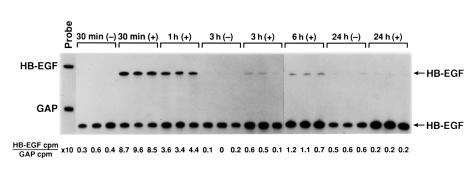
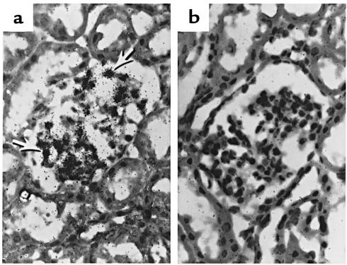
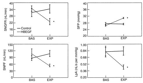
Heparin-binding epidermal growth factor-like growth factor (HB-EGF), a member of the epidermal growth factor (EGF) family, is expressed during inflammatory and pathological conditions. We have cloned the rat HB-EGF and followed the expression of HB-EGF in rat kidneys treated with anti- glomerular basement membrane (anti-GBM) antibody (Ab) to induce glomerulonephritis (GN). We observed glomerular HB-EGF mRNA and protein within 30 minutes of Ab administration and showed by in situ hybridization that glomerular HB-EGF mRNA expression was predominantly in mesangial and epithelial cells. Expression of HB-EGF correlated with the onset of decreased renal function in this model. To test the direct effect of HB-EGF on renal function, we infused the renal cortex with active rHB-EGF, prepared from transfected Drosophila melanogaster cells. This treatment induced a significant decrease in single nephron GFR (SNGFR), single nephron plasma flow, and glomerular ultrafiltration coefficient and an increase in the glomerular capillary hydrostatic pressure gradient. In addition, anti-HB-EGF Ab administered just before anti-GBM Ab blocked the fall in SNGFR and GFR at 90 minutes without any change in the glomerular histologic response. These studies suggest that HB-EGF expressed early in the anti-GBM Ab GN model contributes to the observed acute glomerular hemodynamic alterations.


Figures

Figure 1
Figure 1
Expression of mature, carboxyl his-tagged HB-EGF, purified by nickel column. Cell lysates and purified protein from transformed E. coli were subjected to SDS and 7–20% polyacrylamide gel electrophoresis and stained with Coomassie brilliant blue. The lanes are marked and represent uninduced cell lysates, induced cell lysates 4 hours by IPTG, 1 μg affinity-purified HB-EGF (9.5 kDa), and markers.
Figure 2
Figure 2
Expression of HB-EGF in D. melanogaster cells (SC2). Western blot demonstrated that rabbit anti–HB-EGF Ab generated against E. coli–derived HB-EGF recognized glycosylated 21-kDa HB-EGF in the supernatant of the sense (left lane), but not the mock (right lane), transfectants. The molecular weight was higher than recombinant HB-EGF expressed in E. coli due to the glycosylation process.
Figure 3
Figure 3
Functional analysis of SC2-derived HB-EGF by cell proliferation assay [H3]thymidine incorporation in AKR-2B cells stimulated with recombinant HB-EGF. Two different clones of histidine-fused rHB-EGF (nos. 2 and 3) were functionally active. Data presented are means ± SD of triplicate determinations.
Figure 4
Figure 4
RNase protection assay analysis of mRNA for HB-EGF and GAPDH in glomeruli after anti-GBM Ab administration. The data are presented as a ratio of specific mRNA/GAPDH mRNA to ensure a constant quantity of RNA for each sample. The protected HB-EGF and GAPDH bands are shorter than the respective probes because the unhybridized polylinker regions in the cRNA probes are digested by RNase.
Figure 5
Figure 5
Expression of HB-EGF protein in glomeruli analyzed by immunoprecipitation and Western blot. A total of 500 μg of total glomerular lysate from each of the samples was used. After immunoprecipitation and electroblotting, biotinylated rabbit anti–HB-EGF Ab was incubated with the membrane, and subsequently alkaline phosphatase–conjugated goat antibiotin was used before color development. The experiment was performed as described in the Methods. A band, about 21 kDa, appeared in the lysates from the rats with anti-GBM GN (+), but not from the controls (–).
Figure 6
Figure 6
(a) In situ hybridization analysis of HB-EGF mRNA in kidney tissue 1 hour after anti-GMB Ab administration. The antisense HB-EGF probe hybridized with glomeruli structures, a less intensive signal, was seen in tubules. Glomerular capillary wall and mesangial cells, but not intraluminal inflammatory cells, were the site of mRNA expression (arrows). (b) No signal was found using a sense HB-EGF probe.
Figure 7
Figure 7
Glomerular hemodynamic effects in control rats (solid lines) and rats infused with HB-EGF (dashed lines). SNGFR, SNPF, and LpA significantly decreased in the HB-EGF group. HB-EGF infusion was associated with a significant decrease in SF. *P < 0.05 versus basal period. BAS, basal. EXP, experimental.

Similar articles

Cited by

References

    1. Wilson, C.B. 1996. The renal response to immunological injury. In Brenner and Rector’s the kidney. 5th edition. B.M. Brenner, editor. W.B. Saunders Co., Philadelphia, PA. 1253–1391.
    1. Harris RC. Growth factors and cytokines in acute renal failure. Adv Ren Replace Ther. 1997;4:43–53. - PubMed
    1. Harris RC, Hoover RL, Jacobson HR, Badr KF. Evidence for glomerular actions of epidermal growth factor in the rat. J Clin Invest. 1988;82:1028–1039. - PMC - PubMed
    1. Feng L, Tang WW, Loskutoff DJ, Wilson CB. Dysfunction of glomerular fibrinolysis in experimental anti-glomerular basement membrane antibody glomerulonephritis. J Am Soc Nephrol. 1993;3:1753–1764. - PubMed
    1. Garcia GE, et al. Expression of heparin-binding EGF (HB-EGF) in glomerulonephritis (GN). Recombinant (r) HB-EGF decreased glomerular filtration rate (GRF) J Am Soc Nephrol. 1994;5:692. (Abstr.)

Publication types

MeSH terms

Substances