. 2000 Feb 1;60(3):722-7.

Aminopeptidase N is a receptor for tumor-homing peptides and a target for inhibiting angiogenesis

Affiliations

Aminopeptidase N is a receptor for tumor-homing peptides and a target for inhibiting angiogenesis

R Pasqualini et al. Cancer Res. .

Abstract

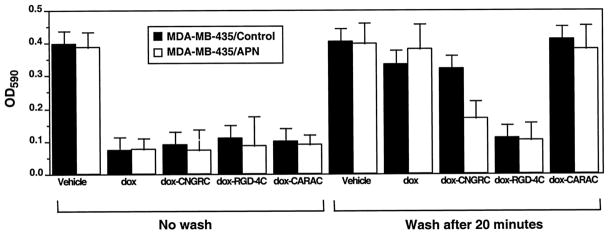
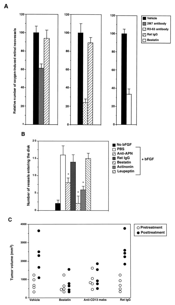
Phage that display a surface peptide with the NGR sequence motif home selectively to tumor vasculature in vivo. A drug coupled to an NGR peptide has more potent antitumor effects than the free drug [W. Arap et al., Science (Washington DC), 279: 377-380, 1998]. We show here that the receptor for the NGR peptides in tumor vasculature is aminopeptidase N (APN; also called CD13). NGR phage specifically bound to immunocaptured APN and to cells engineered to express APN on their surface. Antibodies against APN inhibited in vivo tumor homing by the NGR phage. Immunohistochemical staining showed that APN expression is up-regulated in endothelial cells within mouse and human tumors. In another tissue that undergoes angiogenesis, corpus luteum, blood vessels also expressed APN, but APN was not detected in blood vessels of various other normal tissues stained under the same conditions. APN antagonists specifically inhibited angiogenesis in chorioallantoic membranes and in the retina and suppressed tumor growth. Thus, APN is involved in angiogenesis and can serve as a target for delivering drugs into tumors and for inhibiting angiogenesis.


Figures

Fig. 1
Fig. 1
Specific binding of NGR phage to isolated APN, to APN-transfected cells, and to APN in tumors. A, binding of phage to immunocaptured APN. Phage displaying the RGD or the NGR peptide in different sequence contexts were added at 2 X 10⁹ TUs/well to microtiter wells that had been coated with anti-APN and treated with APN-containing cell extract or with BSA. Data represent the means of bound phage from triplicate wells; bars, SE. B, Inhibition of the binding of NGR phage to APN by soluble NGR peptide. Phage (2 X 10⁹ TUs) were added to microtiter wells, together with 200 μg/well of the indicated peptides, and bound phage were quantitated (means from triplicate wells; bars, SE). C, Binding of NGR phage to APN-transfected cells. APN-transfected and control cells were incubated with CNGRC phage (2 X 10⁹ TUs) and the indicated amounts of the CNGRC or CARAC (control) peptide, and bound phage were quantitated (means from triplicate wells; bars, SE). D, Inhibition of tumor homing of NGR phage by anti-APN. Mice bearing size-matched MDA-MB-435 tumors were coninjected with 109 TUs/mouse of the indicated phage, together with anti-APN IgG or normal rat IgG. The number of phage TUs recovered from the tumors is shown (means; n = 3; bars, SE).
Fig. 2
Fig. 2
An NGR peptide-dox conjugate is selectively toxic to APN-positive cells. APN-transfected and parental MDA-MB-435 cells were exposed either to dox or dox-CNGRC, dox-RGD-4C, or dox-CARAC conjugates. The treatment with the drug was either for the duration of the 24-h incubation (No wash), or the drug was removed by washing cells after 20 min. Incubation continued for another 24 h. Surviving cells were quantitated at the end of the 24-h incubation period. Bars, SE.
Fig. 3
Fig. 3
Immunoperoxidase staining for APN in tumor and normal tissues in mice. A, tumor grown from MDA-MB-435 human breast carcinoma cells in a nude mouse. Antimouse APN shows positive staining in the tumor blood vessels. B, tumor cells and blood vessels in this xenograft are negative with antihuman APN, which does not react with mouse APN. Mouse liver (C) and spleen (D) show no significant staining with the antimouse APN antibody.
Fig. 4
Fig. 4
APN expression in human angiogenesis. Immunoperoxidase staining shows strong APN expression in endothelial cells and the subendothelium of blood vessels in a human breast carcinoma (A) and in corpus luteum undergoing angiogenesis (C). The blood vessels in normal breast tissue are essentially negative (B). D is a confocal immunofluorescence image showing anti-APN staining of a medium-sized vessel in a human carcinoma. APN staining is present both at the endothelial surface and in a subendothelial layer. A–C, X300; D, X500.
Fig. 5
Fig. 5
Inhibition of angiogenesis by APN inhibitors. A, mice developing hypoxia-induced retinal neovasculature were treated i.v. with PBS (Vehicle), antimouse APN antibodies or normal rat IgG (250 μg/mouse), or bestatin (200 μg/mouse). The number of retinal neovessels in mice treated with PBS was set at 100%. Data represent means; n = 3; bars, SE. The reduction in blood vessel number was statistically significant for the anti-APN antibodies and bestatin (P < 0.01). B, CAMs were treated with PBS, bFGF, or bFGF together with anti-APN R3-63, normal rat IgG, bestatin, actinonin, or leupeptin. There were significantly fewer vessels in the CAMs treated with the APN inhibitors, whereas the serine protease inhibitor, leupeptin, had no effect. Columns, means; n = 8; bars, SE. *, t test, P < 0.05 relative to controls. C, mice bearing size-matched MDA-MB-435-derived breast carcinoma xenografts were divided into four groups of five animals and treated with DMEM, bestatin (250 μg/mouse), a mixture of R3-63 and 2M-7 anti- bodies, or normal rat IgG (125 μg/mouse) given i.p. once a week for 3 weeks. Tumor volumes at the start of the experiment (Pretreatment) and 3 weeks later (Posttreatment) are shown. Similar results were observed in two independent experiments. The tumors in the groups treated with bestatin and anti-APN had a significantly smaller volume posttreatment than the control groups (t test, P < 0.05).

References

    1. Folkman J. Angiogenesis in cancer, vascular, rheumatoid and other disease. Nat Med. 1995;1:27–31. - PubMed
    1. Hanahan D, Folkman J. Patterns and emerging mechanisms of the angiogenic switch during tumorigenesis. Cell. 1996;86:353–364. - PubMed
    1. Risau W, Flamme I. Vasculogenesis. Annu Rev Cell Dev Biol. 1995;11:73–91. - PubMed
    1. Friedlander M, Brooks PC, Shaffer RW, Kincaid CM, Varner JA, Cheresh DA. Definition of two angiogenic pathways by distinct αv integrins. Science (Washington DC) 1995;270:1500–1502. - PubMed
    1. Hammes HP, Brownlee M, Jonczyk A, Sutter A, Preissner KT. Subcutaneous injection of a cyclic peptide antagonist of vitronectin receptor-type integrins inhibits retinal neovascularization. Nat Med. 1996;2:529–533. - PubMed

Publication types

LinkOut - more resources