. 2000 Feb;66(2):356-67.
doi: 10.1086/302751.

Exon skipping in IVD RNA processing in isovaleric acidemia caused by point mutations in the coding region of the IVD gene

Affiliations

Exon skipping in IVD RNA processing in isovaleric acidemia caused by point mutations in the coding region of the IVD gene

J Vockley et al. Am J Hum Genet. 2000 Feb.

Abstract

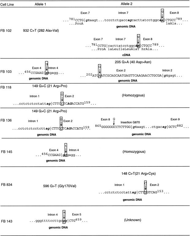
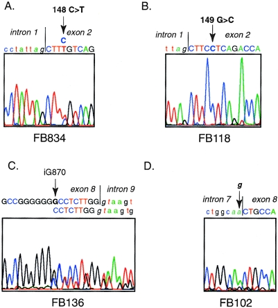
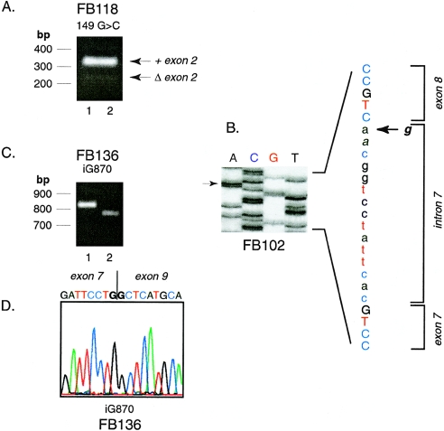
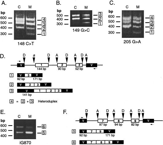
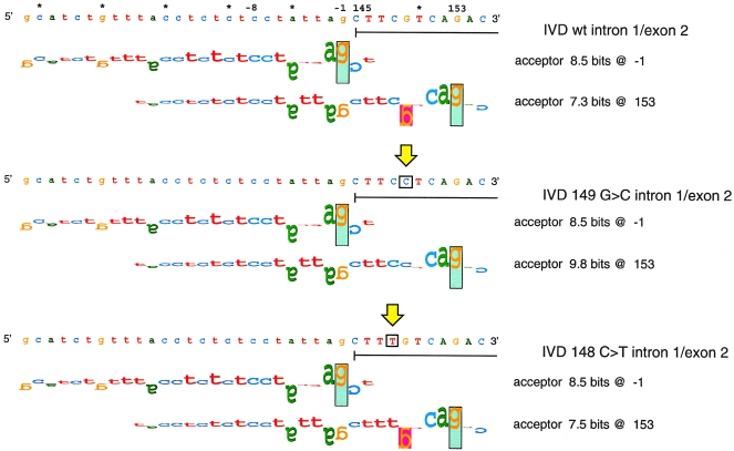
Isovaleric acidemia (IVA) is a recessive disorder caused by a deficiency of isovaleryl-CoA dehydrogenase (IVD). We have reported elsewhere nine point mutations in the IVD gene in fibroblasts of patients with IVA, which lead to abnormalities in IVD protein processing and activity. In this report, we describe eight IVD gene mutations identified in seven IVA patients that result in abnormal splicing of IVD RNA. Four mutations in the coding region lead to aberrantly spliced mRNA species in patient fibroblasts. Three of these are amino acid altering point mutations, whereas one is a single-base insertion that leads to a shift in the reading frame of the mRNA. Two of the coding mutations strengthen pre-existing cryptic splice acceptors adjacent to the natural splice junctions and apparently interfere with exon recognition, resulting in exon skipping. This mechanism for missplicing has not been reported elsewhere. Four other mutations alter either the conserved gt or ag dinucleotide splice sites in the IVD gene. Exon skipping and cryptic splicing were confirmed by transfection of these mutations into a Cos-7 cell line model splicing system. Several of the mutations were predicted by individual information analysis to inactivate or significantly weaken adjacent donor or acceptor sites. The high frequency of splicing mutations identified in these patients is unusual, as is the finding of missplicing associated with missense mutations in exons. These results may lead to a better understanding of the phenotypic complexity of IVA, as well as provide insight into those factors important in defining intron/exon boundaries in vivo.


Figures

Figure  1
Figure 1
Summary of mutations in the IVD gene leading to abnormal splicing of IVD RNA. IVD sequences, including intron and exon boundaries, were amplified from genomic DNA isolated from cell lines from patients with IVA and shown to be deficient in IVD enzymatic activity. Mutations identified are listed. For FB102, the effect of the intron 7 mutation on cDNA sequence is also shown. FB118 and FB145 were homozygous for their respective mutations. Only one mutation could be identified in FB143, despite complete sequencing of all exons and extensive sequencing of the intron/exon boundaries. The remaining mutation in the IVD gene responsible for this patient’s IVA is unknown.
Figure  2
Figure 2
Sequencing of DNA fragments amplified from IVD genomic DNA from IVA fibroblasts. A, Direct sequence of amplified genomic DNA from FB834, showing a 148C→T mutation. The patient is heterozygous at this position. B, 149G→C mutation in FB118 in products amplified from genomic DNA and sequenced directly. The patient is homozygous for this mutation, which was also identified in FB136. Control experiments confirmed that this was not related to contamination of PCR reactions with previously amplified template. C, Insertion of a G residue in genomic DNA from FB136 in an area that normally contains six Gs from cDNA positions 865–870. Direct sequence from amplified genomic DNA is shown. Because the patient is heterozygous for this mutation, a double sequencing pattern appears after the inserted nucleotide. This finding was confirmed in the reverse direction and by sequencing of subclones of the PCR amplified genomic DNA from this region (not shown). D, G→A point mutation of the intron 7 splice acceptor site in FB102 genomic DNA, identified by direct sequencing of amplified DNA. The patient is heterozygous at this location. The mutant A allele is easily visualized in the forward direction shown, whereas the wild-type G signal is more evident in the reverse direction (not shown).
Figure  3
Figure 3
Identification of exon skipping in mRNA from IVA fibroblasts. A, Agarose gel of DNA fragments amplified from cDNA from FB118. The 5′ amplification primer was in the 5′ untranslated region of IVD (position −20 to −1) and the 3′ amplification primer was from position 265–286, spanning exons 1–3. Arrows mark the predicted size of a normal fragment (306 bp) and one with a deletion of exon 2 (90 bp). The migration positions of 100 bp molecular mass standards are shown. B, Manual DNA sequencing of a subcloned DNA fragment amplified from FB102 cDNA, including exons 7 and 8. The arrows show the position of the ga mutation in the intron 7 splice acceptor site. Sequences corresponding to exon 7, intron 7, and exon 8 are marked. C and D, Analysis of subclones of amplified products from FB136 cDNA, spanning exons 3–11 (5′ primer, IVD coding nucleotides 318–347; 3′ primer, IVD coding nucleotides 1122–1138; anticipated size 820 bp). Two different-sized inserts are obtained after subcloning, as seen on agarose gel electrophoresis (C). The sequence of the insert from lane 1 is normal (not shown). The sequence of the insert from lane 2 shows a deletion of exon 8 (D).
Figure  4
Figure 4
Use of an exon-trapping system to examine the effects of IVD exon mutations on RNA splicing. IVD genomic DNA fragments, containing control or mutation sequences, spanning exon 1 to intron 3 (AD) or intron 7 to intron 9 (EF) were cloned into the exon-trapping vector pSPL3. Inserts for all clones were sequenced to confirm that no PCR errors were present. The vector was transiently transfected into COS-7 cells, and total RNA from transfected cells was used as template for RT-PCR, with vector-specific primers. Amplified products were separated by electrophoresis on agarose gels and visualized with ethidium bromide staining. The number of products listed below the vector maps correspond to the numbering of the bands on the gel photo. Lanes containing PCR products amplified from cDNA of cells transfected with control and mutant IVD sequences are marked as “C” and “M,” respectively. The mutation being analyzed is listed underneath, and the migration of molecular mass markers is indicated to the left of each gel photograph. A, 148C→T; B, 149G→C; C, 205G→A. D and F, Map of amplified products identified by sequencing subcloned products obtained with normal and mutant IVD exon-trapping experiments. The test IVD exon-trapping vector plus insert is depicted on the top line. IVD exons are shown as white boxes and exon sizes are as labeled. Vector exon sequences (V) are denoted by black boxes and intron sequences by a line. Donor and acceptor sites are indicated by D and A, respectively. The horizontal arrows indicate the position of PCR primers. D and F, Exon 2 splicing test vector. Cryptic acceptor site in vector is indicated by “a.” E, iG870 = insertion of G at position 870 in the IVD coding region. F, Exon 8 splicing test vector.
Figure  5
Figure 5
Walker diagrams of IVD exon 2 splice acceptor sites. Splice sites are shown by walkers (Schneider 1997b), in which the height of each letter is the contribution of that base to the total conservation of the site (in bits). The upper bound of the vertical rectangles is at +2 bits, and their lower bound is at −3 bits. Letters that are upside down represent negative contributions. The top panel depicts the natural splice site one nucleotide upstream of exon 2 and a cryptic acceptor site at position 153. Neither cryptic site is used. The vertical arrows indicate the locations of mutations in the sequence. The middle panel corresponds to the G→C change at position 149, and the lower panel to C→T at position 148. The mutations alter the Ri value of the cryptic acceptor at position 153.
Figure  6
Figure 6
Polymorphic repeats in intron 1 of the IVD gene. A, Sequence of three, two, and one repeat alleles in intron 1, beginning 25 bp from the junction of exon 1 and intron 1. B, Amplification of intron 1 repeats from fibroblast cDNA. Fragments corresponding to each number of repeats are shown.

Similar articles

Cited by

References

Electronic-Database Information

    1. GenBank, http://www.ncbi.nlm.nih.gov/Genbank (accession numbers AF191214 to AF191218)
    1. Laboratory of Computational and Experimental Biology at the National Institutes of Health, http://www.lecb.ncifcrf.gov/~toms/ (for the Scan program)
    1. Online Mendelian Inheritance in Man (OMIM), http://www.ncbi.nlm.nih.gov/Omim (for IVA [MIM 243500])

References

    1. Berg MA, Guevara-Aguirre J, Rosenbloom AL, Rosenfeld RG, Francke U (1992) Mutation creating a new splice site in the growth hormone receptor genes of 37 Ecuadorean patients with Laron syndrome. Hum Mutat 1:24–32 - PubMed
    1. Crane F, Beinert H (1955) On the mechanism of dehydrogenation of fatty acyl derivatives of coenzyme A: part II. The electron-transferring flavoproteins. J Biol Chem 218:717–731 - PubMed
    1. Dietz HC, Kendzior RJ Jr. (1994) Maintenance of an open reading frame as an additional level of scrutiny during splice site selection. Nat Genet 8:183–188 - PubMed
    1. Dietz HC, Valle D, Francomano CA, Kendzior RJ Jr., Pyeritz RE, Cutting GR (1993) The skipping of constitutive exons in vivo induced by nonsense mutations. Science 259:680–683 - PubMed
    1. Graveley BR, Hertel KJ, Maniatis T (1998) A systematic analysis of the factors that determine the strength of pre-mRNA splicing enhancers. EMBO J 17:6747–6756 - PMC - PubMed

Publication types

MeSH terms