

The X chromosome frequently lags behind in female lymphocyte anaphase
- PMID: 10677326
- PMCID: PMC1288119
- DOI: 10.1086/302769
The X chromosome frequently lags behind in female lymphocyte anaphase
Abstract
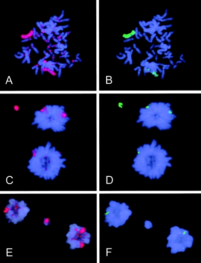
Pancentromeric FISH and X-chromosome painting were used to characterize anaphase aberrations in 2,048 cultured lymphocytes from a healthy 62-year-old woman. Of 163 aberrant anaphases, 66.9% contained either chromosomes or their fragments that lagged behind. Characterization of 200 laggards showed that 49% were autosomes, 33. 5% were autosomal fragments, and 17.5% were X chromosomes. The X chromosome represented one-fourth of all lagging chromosomes and was involved much more often than would be expected by chance (1/23). Labeling of the late-replicating inactive X chromosome with 5-bromo-2'-deoxyuridine revealed that both X homologues contributed equally to the laggards. Among 200 micronuclei examined from interphase cells, the proportion of the X chromosome (31%) and autosomal fragments (50%) was higher than among anaphase laggards, whereas autosomes were involved less often (19%). These findings may reflect either selection or the fact that lagging autosomes, which were more proximal to the poles than were lagging X chromosomes, were more frequently included within the main nucleus. Our results suggest that the well-known high micronucleation and loss of the X chromosome in women's lymphocytes is the result of frequent distal lagging behind in anaphase and effective micronucleation of this chromosome. This lagging appears to affect the inactive and active X chromosomes equally.
Figures
References
Electronic-Database Information
-
- Center for Molecular Genetics and Toxicology, http://www.swan.ac.uk/cget/ejgt/article4.htm (for Stopper [] reference article)
References
-
- Abruzzo MA, Mayer M, Jacobs PA (1985) Aging and aneuploidy: evidence for the preferential involvement of the inactive X chromosome. Cytogenet Cell Genet 39:275–278 - PubMed
-
- Catalán J, Autio K, Wessman M, Lindholm C, Knuutila S, Sorsa M, Norppa H (1995) Age-associated micronuclei containing centromeres and the X chromosome in lymphocytes of women. Cytogenet Cell Genet 68:11–16 - PubMed
-
- Fitzgerald PH, McEwan CM (1977) Total aneuploidy and age-related sex chromosome aneuploidy in cultured lymphocytes of normal men and women. Hum Genet 39:329–337 - PubMed
-
- Ford JH, Congedi MM (1987) Rapid induction of anaphase in competent cells by hypotonic treatment. Cytobios 51:183–192 - PubMed
Publication types
MeSH terms
Substances
LinkOut - more resources
Full Text Sources
