

Review
doi: 10.1136/heart.83.3.361.
The pathophysiology of acute coronary syndromes
Affiliations
- PMID: 10677422
- PMCID: PMC1729334
- DOI: 10.1136/heart.83.3.361
Item in Clipboard
Review
The pathophysiology of acute coronary syndromes
Heart.
2000 Mar.
No abstract available
Figures

The established stable plaque. In this cross section of a human coronary artery there is an established fibrolipid plaque with a core of lipid. The lipid core is separated from the lumen by the plaque cap. The plaque only occupies part of the circumference of the artery, leaving a segment of normal arterial wall.
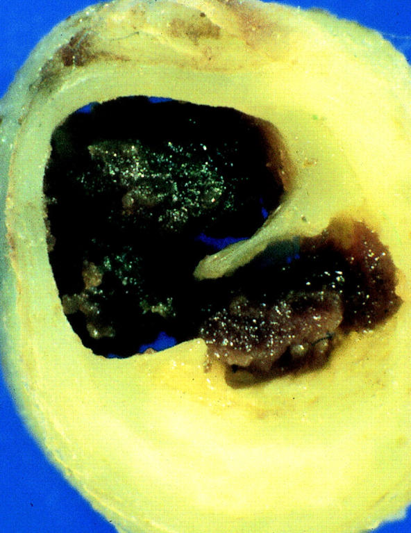
Thrombosis caused by erosion. This human coronary artery is largely occluded by a mass of thrombus which is adherent to the surface of a plaque. The plaque itself is intact.
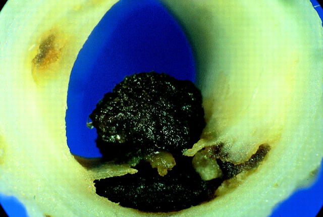
Thrombosis caused by disruption. The cap of a plaque has torn and projects up into the lumen. Thrombus has formed within the original lipid core from where it projects into, but does not totally occlude, the lumen. This is the typical lesion of unstable angina.
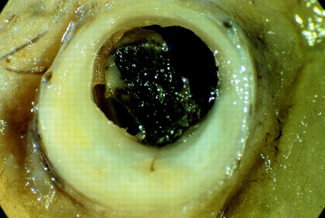
Thrombosis caused by disruption. The cap of the plaque has torn and thrombus within the lipid core extends into and occludes the lumen. This is the typical lesion of acute myocardial infarction.
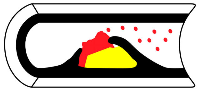
Platelet embolisation. Any thrombus (red) which protrudes into the arterial lumen but does not occlude has the surface covered by a layer of activated platelets strongly expressing the IIb/IIIa receptor. Clumps of these platelets are swept down into the myocardium vascular bed.
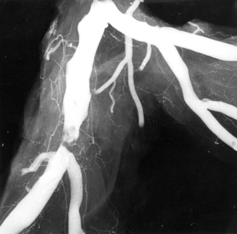
Angiogram of plaque disruption. In this postmortem angiogram there is a typical type II eccentric ragged stenosis with an overlying intraluminal filling defect indicating thrombus over the plaque.
References
Publication types
MeSH terms
LinkOut - more resources
Full Text Sources
Other Literature Sources
Medical