. 2000 Feb;122(2):319-26.
doi: 10.1104/pp.122.2.319.

Amino acid transporters are localized to transfer cells of developing pea seeds

Affiliations

Amino acid transporters are localized to transfer cells of developing pea seeds

M Tegeder et al. Plant Physiol. 2000 Feb.

Abstract

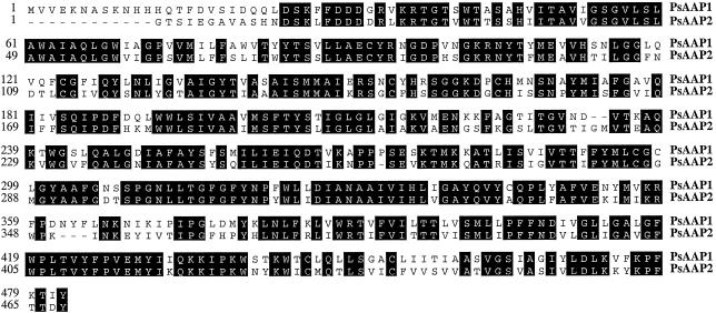
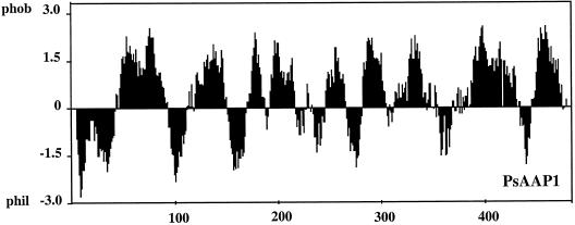
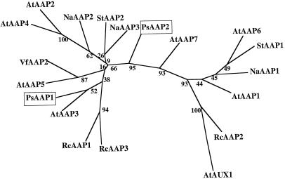
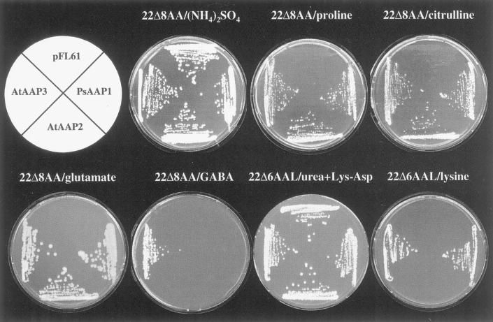
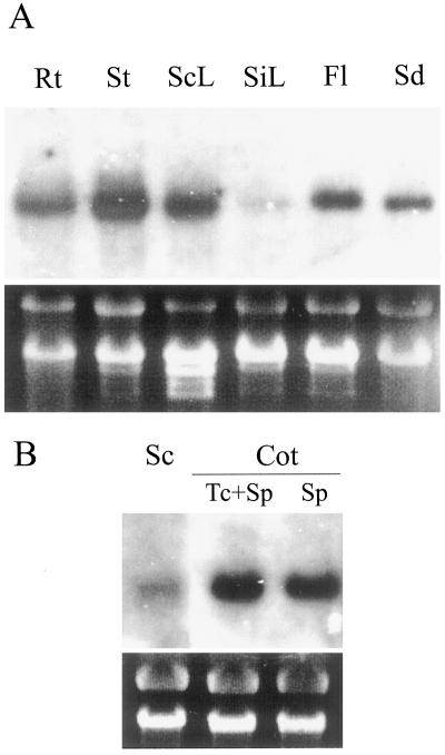
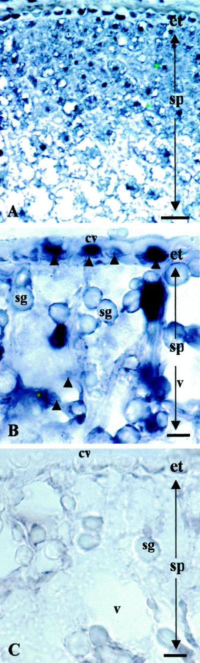
To determine the nature and cellular localization of amino acid transport in pea seeds, two cDNA clones belonging to the AAP family of H(+)/amino acid co-transporters (PsAAP1 and PsAAP2) were isolated from a cotyledon cDNA library of pea (Pisum sativum L.). Functional expression in the yeast amino acid uptake mutants 22Delta6AAL and 22Delta8AA showed that PsAAP1 mediates transport of neutral, acidic, and basic amino acids. RNA-blot analyses showed that PsAAP1 is expressed in seeds and vegetative organs, including amino acid sinks and sources, whereas PsAAP2 could not be detected. For developing seeds, transcripts of PsAAP1 were detected in coats and cotyledons, with seed coats giving a weak signal. In cotyledons, expression was highest in epidermal-transfer-cell-enriched tissue. RNA in situ hybridization analysis showed that PsAAP1 was predominantly present in epidermal transfer cells forming the outer surface of cotyledons, which abuts the seed coats. Overall, our observations suggest that this transporter, which is localized in transfer cells of cotyledons, might play a role in the uptake of the full spectrum of amino acids released from seed coats.


Figures

Figure 1
Figure 1
Amino acid sequence alignment of amino acid transporters PsAAP1 and PsAAP2 from pea.
Figure 2
Figure 2
Prediction of putative membrane-spanning regions in PsAAP1 using LASERGENE software (DNASTAR, Madison, WI).
Figure 3
Figure 3
Computer-aided analysis of homologies between PsAAP1 and PsAAP2 and related proteins. The analysis was performed using PHYLIP (J. Felsenstein, 1995, Phylogenetic Interference Package, version 3.5, distributed by the author: Department of Genetics, University of Washington, Seattle, WA) with aligned sequences of PsAAP1 and PsAAP2 and amino acid transporters from Arabidopsis (AtAAPs), S. tuberosum (StAAPs), R. communis (RcAAPs; RcAAP3-accession no. AJ132228) (Fischer et al., 1998), N. alata (NaAAPs; Schulze et al., 1999), and V. faba (VfAAP2, accession no. Y09591). The numbers indicate the occurrence of a given branch in 100 bootstrap replicates of the given data set. AtAUX1 was used as the outgroup.
Figure 4
Figure 4
Complementation of growth of yeast strains 22Δ8AA and 22Δ6AAL, which are deficient in the uptake of amino acids, by PsAAP1, AtAAP2, and AtAAP3. Growth of 22Δ8AA and 22Δ6AAL expressing PsAAP1, AtAAP2, and AtAAP3 and the strains transformed with the vector pFL61 is shown. For selection, the medium was supplemented with Pro, citrulline, Gaba, Glu (22Δ8AA), or Lys (22Δ6AAL) as the sole nitrogen source. For non-selective conditions, the medium contained either ammonium sulfate (22Δ8AA) or urea and the dipeptide Lys-Asp as the sole nitrogen source (22Δ6AAL).
Figure 5
Figure 5
Profiles of organ expression of PsAAP1. Gel blots of total RNA (20 μg per lane) were hybridized with 32P-labeled 3′-untranslated region cDNA probe of PsAAP1. Ethidium bromide staining of rRNA is shown at bottom. A, Northern-blot analyses of RNA extracted from different pea organs. Rt, Roots; St, stems; ScL, source leaves; SiL, sink leaves; Fl, flowers; and Sd, developing seeds. B, Tissue-specific expression of PsAAP1 in developing pea seeds. Northern-blot analyses of RNA extracted from seed coat (Sc) and from cotyledon (Cot) epidermal-transfer-cell-enriched (Tc+Sp) and cotyledon storage parenchyma (Sp) tissues.
Figure 6
Figure 6
Light micrographs illustrating in situ hybridization of DIG-labeled PsAAP1 antisense (A and B) and sense (C) riboprobes to transverse sections of developing pea cotyledons. Probe binding in the cytoplasm of the epidermal transfer cells and the storage parenchyma cells is evident as a strong blue color indicated by darts in B (compare with the sense control, C). The dense color reactant in the epidermal transfer cells and five to six rows of storage parenchyma cells inward of the epidermis declines through subsequent consecutive rows of storage parenchyma cells (A). In A, the bar = 100 μm (×120); in B and C, the bar = 30 μm (×300). cw, Cell wall; et, epidermal transfer cell; sg, starch granule; sp, storage parenchyma cell; and v, vacuole.

References

    1. Bick JA, Neelam A, Hall JL, Williams LE. Amino acid carriers of Ricinus communis expressed during seedling development: molecular cloning and expression analysis of two putative amino acid transporters, RcAAP1 and RcAAP2. Plant Mol Biol. 1998;36:377–385. - PubMed
    1. Borstlap AC, Schuurmans J. Kinetics of l-valine uptake in tobacco leaf discs: comparison of wild type, the digenic mutant Valr-2 and its monogenic derivatives. Planta. 1988;176:42–50. - PubMed
    1. Bush DR, Langston-Unkefer PJ. Amino acid transport into membrane vesicles isolated from zucchini. Plant Physiol. 1988;88:487–490. - PMC - PubMed
    1. De Jong A, Koerselman-Kooij JW, Schuurmans JAMJ, Borstlap AC. The mechanisms of amino acid efflux from seed coats of developing pea seeds as revealed by uptake experiments. Plant Physiol. 1997;114:731–736. - PMC - PubMed
    1. De Jong A, Wolswinkel P. Differences in release of endogenous sugars and amino acids from attached and detached seed coats of developing pea seeds. Physiol Plant. 1995;94:78–86.

Publication types

LinkOut - more resources