

Expression and localization of nitrilase during symptom development of the clubroot disease in Arabidopsis
- PMID: 10677430
- PMCID: PMC58874
- DOI: 10.1104/pp.122.2.369
Expression and localization of nitrilase during symptom development of the clubroot disease in Arabidopsis
Abstract
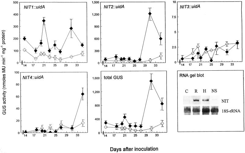
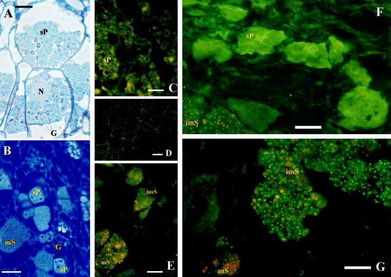
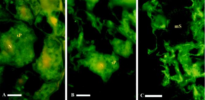
The expression of nitrilase in Arabidopsis during the development of the clubroot disease caused by the obligate biotroph Plasmodiophora brassicae was investigated. A time course study showed that only during the exponential growth phase of the clubs was nitrilase prominently enhanced in infected roots compared with controls. NIT1 and NIT2 are the nitrilase isoforms predominantly expressed in clubroot tissue, as shown by investigating promoter-beta-glucuronidase fusions of each. Two peaks of beta-glucuronidase activity were visible: an earlier peak (21 d post inoculation) consisting only of the expression of NIT1, and a second peak at about 32 d post inoculation, which predominantly consisted of NIT2 expression. Using a polyclonal antibody against nitrilase, it was shown that the protein was mainly found in infected cells containing sporulating plasmodia, whereas in cells of healthy roots and in uninfected cells of inoculated roots only a few immunosignals were detected. To determine which effect a missing nitrilase isoform might have on symptom development, the P. brassicae infection in a nitrilase mutant (nit1-3) of Arabidopsis was investigated. As a comparison, transgenic plants overexpressing NIT2 under the control of the cauliflower mosaic virus 35S promoter were studied. Root galls were smaller in nit1-3 plants compared with the wild type. The phenotype of smaller clubs in the mutant was correlated with a lower free indole-3-acetic acid content in the clubs compared with the wild type. Overexpression of nitrilase did not result in larger clubs compared with the wild type. The putative role of nitrilase and auxins during symptom development is discussed.
Figures
Similar articles
-
Structural analysis of the nit2/nit1/nit3 gene cluster encoding nitrilases, enzymes catalyzing the terminal activation step in indole-acetic acid biosynthesis in Arabidopsis thaliana.Plant Mol Biol. 1998 Jan;36(1):89-99. doi: 10.1023/a:1005998918418. Plant Mol Biol. 1998. PMID: 9484465
-
Enzymatic characterization of the recombinant Arabidopsis thaliana nitrilase subfamily encoded by the NIT2/NIT1/NIT3-gene cluster.Planta. 2001 Mar;212(4):508-16. doi: 10.1007/s004250000420. Planta. 2001. PMID: 11525507
-
A role for nitrilase 3 in the regulation of root morphology in sulphur-starving Arabidopsis thaliana.Plant J. 2002 Apr;30(1):95-106. doi: 10.1046/j.1365-313x.2002.01271.x. Plant J. 2002. PMID: 11967096
-
Plasmodiophora brassicae: a review of an emerging pathogen of the Canadian canola (Brassica napus) crop.Mol Plant Pathol. 2012 Feb;13(2):105-13. doi: 10.1111/j.1364-3703.2011.00729.x. Epub 2011 Jun 1. Mol Plant Pathol. 2012. PMID: 21726396 Free PMC article. Review.
-
Getting to the root of a club - Understanding developmental manipulation by the clubroot pathogen.Semin Cell Dev Biol. 2023 Oct-Nov;148-149:22-32. doi: 10.1016/j.semcdb.2023.02.005. Epub 2023 Feb 13. Semin Cell Dev Biol. 2023. PMID: 36792438 Review.
Cited by
-
Plant tumors: a hundred years of study.Planta. 2020 Mar 18;251(4):82. doi: 10.1007/s00425-020-03375-5. Planta. 2020. PMID: 32189080 Review.
-
Dynamic Modeling of Indole Glucosinolate Hydrolysis and Its Impact on Auxin Signaling.Front Plant Sci. 2018 Apr 26;9:550. doi: 10.3389/fpls.2018.00550. eCollection 2018. Front Plant Sci. 2018. PMID: 29755493 Free PMC article.
-
Root Transcriptome and Metabolome Profiling Reveal Key Phytohormone-Related Genes and Pathways Involved Clubroot Resistance in Brassica rapa L.Front Plant Sci. 2021 Dec 15;12:759623. doi: 10.3389/fpls.2021.759623. eCollection 2021. Front Plant Sci. 2021. PMID: 34975941 Free PMC article.
-
Transcriptomic response in symptomless roots of clubroot infected kohlrabi (Brassica oleracea var. gongylodes) mirrors resistant plants.BMC Plant Biol. 2019 Jul 1;19(1):288. doi: 10.1186/s12870-019-1902-z. BMC Plant Biol. 2019. PMID: 31262271 Free PMC article.
-
Mining of Brassica-Specific Genes (BSGs) and Their Induction in Different Developmental Stages and under Plasmodiophora brassicae Stress in Brassica rapa.Int J Mol Sci. 2018 Jul 16;19(7):2064. doi: 10.3390/ijms19072064. Int J Mol Sci. 2018. PMID: 30012965 Free PMC article.
References
-
- Allard R. The functional analysis of plant nitrilases in transgenic plants. PhD thesis. Germany: Göttingen; 1995. . ISBN 3–89588–172–4.
-
- Bartling D, Seedorf M, Mithöfer A, Weiler EW. Cloning and expression of an Arabidopsis nitrilase which can convert indole-3-acetonitrile to the plant hormone indole-3-acetic acid. Eur J Biochem. 1992;205:417–424. - PubMed
-
- Baskin TI, Busby CH, Fowke LC, Sammut M, Gabler F. Improvements in immunostaining samples embedded in methacrylate: localisation of microtubules and other antigens throughout developing plants of diverse taxa. Planta. 1992;187:405–413. - PubMed
Publication types
MeSH terms
Substances
LinkOut - more resources
Full Text Sources
Other Literature Sources
Molecular Biology Databases
Research Materials
Miscellaneous
