. 2000 Feb 15;97(4):1444-9.
doi: 10.1073/pnas.97.4.1444.

The subcellular localization of acetyl-CoA carboxylase 2

Affiliations

The subcellular localization of acetyl-CoA carboxylase 2

L Abu-Elheiga et al. Proc Natl Acad Sci U S A. .

Abstract

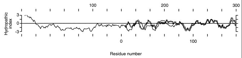
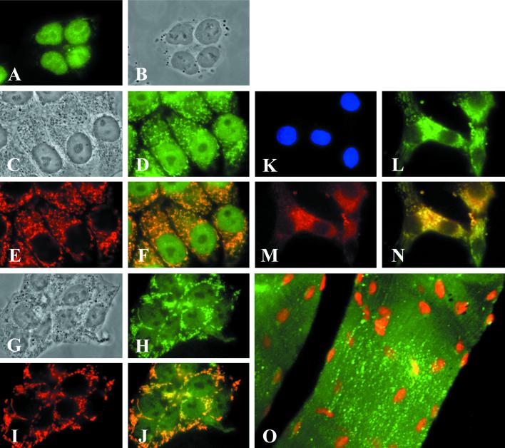
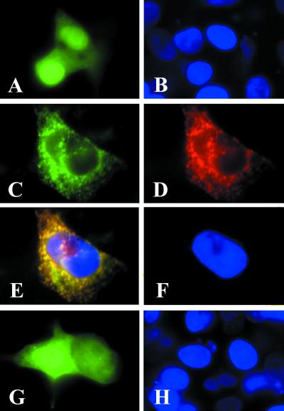
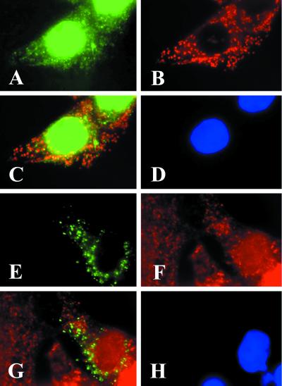
Animals, including humans, express two isoforms of acetyl-CoA carboxylase (EC ), ACC1 (M(r) = 265 kDa) and ACC2 (M(r) = 280 kDa). The predicted amino acid sequence of ACC2 contains an additional 136 aa relative to ACC1, 114 of which constitute the unique N-terminal sequence of ACC2. The hydropathic profiles of the two ACC isoforms generally are comparable, except for the unique N-terminal sequence in ACC2. The sequence of amino acid residues 1-20 of ACC2 is highly hydrophobic, suggesting that it is a leader sequence that targets ACC2 for insertion into membranes. The subcellular localization of ACC2 in mammalian cells was determined by performing immunofluorescence microscopic analysis using affinity-purified anti-ACC2-specific antibodies and transient expression of the green fluorescent protein fused to the C terminus of the N-terminal sequences of ACC1 and ACC2. These analyses demonstrated that ACC1 is a cytosolic protein and that ACC2 was associated with the mitochondria, a finding that was confirmed further by the immunocolocalization of a known human mitochondria-specific protein and the carnitine palmitoyltransferase 1. Based on analyses of the fusion proteins of ACC-green fluorescent protein, we concluded that the N-terminal sequences of ACC2 are responsible for mitochondrial targeting of ACC2. The association of ACC2 with the mitochondria is consistent with the hypothesis that ACC2 is involved in the regulation of mitochondrial fatty acid oxidation through the inhibition of carnitine palmitoyltransferase 1 by its product malonyl-CoA.


Figures

Figure 1
Figure 1
Hydropathic profiles of the 300-residue N terminus of ACC2 (solid line) and the 160-residue N terminus of ACC1 (dotted line) according to Kyte and Doolittle (28) free-energy values. The sequence alignment was used as a basis for aligning the hydrophobicity plots of the N termini of ACC2 and ACC1.
Figure 2
Figure 2
Determination of the mitochondrial localization of ACC2 by fluorescence microscopic analysis. HepG2 cells (A−F), T47D cells (G−J), and neonatal rat cardiomyocytes (K−N) were plated separately at a density of 2 × 10⁵ on glass coverslips coated with polyamino acids. After 48 h of incubation in medium containing 10% bovine serum, the cells were fixed in 3.7% formaldehyde and then immunostained with preimmune serum (A) or ACC2 antibodies and mAbs raised against mitochondria-specific protein as a marker (D, E, H, L, and M). The cells were visualized by reacting them with the goat anti-mouse IgG-TXRD conjugate for the mitochondrial proteins (red) or with the goat anti-rabbit IgG-FITC conjugate (green) for ACC2. (B, C, and G) The images were visualized by phase-contrast microscopy and clearly show the nuclei and cytoplasm of the cells. (F, G, and N) Composite images, respectively, of D and E, H and I, and L and M. The overlapping regions in F, G, and N are yellow. (O) Mouse skeletal muscle tissue that was immunostained with ACC2 antibodies and was visualized by reacting it with the goat anti-rabbit IgG-FITC conjugate (green). The nuclei of the skeletal muscle cells were stained with 0.5 μg of propidium iodide. (K) Neonatal rat cardiomyocytes were counterstained with DAPI.
Figure 3
Figure 3
The ACC2 N terminus directs the GFP to the mitochondria. Neonatal rat cardiomyocytes were plated at a density of 2 × 10⁵ on glass coverslips as described in the legend to Fig. 2. The cells were transfected with 1 μg/ml pEGFP-N1 (A and B), pEGFP-N1-ACC2-N (C and D), or pEGFP-N1-ACC1-N (G and H) by using the transfection reagent Lipofectamine according to the manufacturer's protocol. After 48 h of transfection, the cells were examined by fluorescence microscopy to verify expression of the GFP fusion protein (green). Cells that were transfected with pEGFP-N1-ACC2-N were immunostained with ACC2 antibodies and visualized by reacting them with the goat anti-rabbit IgG-TXRD conjugate (D). (E) A composite image of C and D; the overlapping regions in E are yellow. The cells were counterstained with DAPI (blue).
Figure 4
Figure 4
CPT1 and the GFP1-N1-ACC2-N fusion protein are colocalized on the mitochondria. HepG2 cells were plated at a density of 2 × 10⁵ on glass coverslips for immunostaining and transfection as described in the legends to Figs. 2 and 3. The cells were immunostained with CPT1 antibodies that were detected by reacting the cells with the goat anti-rabbit IgG-FITC conjugate (A) and with mAbs raised against the mitochondrial protein and were visualized by reacting them with the goat anti-mouse IgG-TXRD conjugate (B). (C) A composite image of A and B; the overlapping regions in C are yellow. In the transfection experiments, the cells were transfected with pEGFP-N1-ACC2-N, as described in Materials and Methods, and were immunostained with CPT1 antibodies. The expressed GFP-N1-ACC2-N is green (E). Immunostaining of CPT1 was detected by reacting the cells with the goat anti-rabbit IgG-TXRD conjugate (F). (G) A composite image of E and F; the overlapping regions in G are yellow. The cell nuclei were counterstained with DAPI (blue in D and H).

References

    1. Wakil S J. Am J Chem Soc. 1958;80:6465.
    1. Nugteren D H. Biochim Biophys Acta. 1965;106:280–290. - PubMed
    1. Hopwood D A, Sherman D H. Annu Rev Genet. 1990;24:37–66. - PubMed
    1. Thampy K G. J Biol Chem. 1989;264:17631–17634. - PubMed
    1. Bianchi A, Evans J L, Iverson A J, Nordlund A-C, Watts T D, Witters L A. J Biol Chem. 1990;265:1502–1509. - PubMed

Publication types