. 2000 Feb 15;97(4):1450-5.
doi: 10.1073/pnas.040548197.

A structural basis for integrin activation by the cytoplasmic tail of the alpha IIb-subunit

Affiliations

A structural basis for integrin activation by the cytoplasmic tail of the alpha IIb-subunit

O Vinogradova et al. Proc Natl Acad Sci U S A. .

Abstract

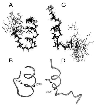
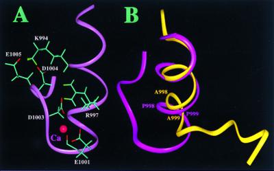
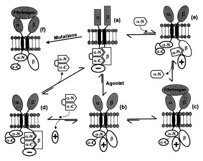
A key step in the activation of heterodimeric integrin adhesion receptors is the transmission of an agonist-induced cellular signal from the short alpha- and/or beta-cytoplasmic tails to the extracellular domains of the receptor. The structural details of how the cytoplasmic tails mediate such an inside-out signaling process remain unclear. We report herein the NMR structures of a membrane-anchored cytoplasmic tail of the alpha(IIb)-subunit and of a mutant alpha(IIb)-cytoplasmic tail that renders platelet integrin alpha(IIb)beta(3) constitutively active. The structure of the wild-type alpha(IIb)-cytoplasmic tail reveals a "closed" conformation where the highly conserved N-terminal membrane-proximal region forms an alpha-helix followed by a turn, and the acidic C-terminal loop interacts with the N-terminal helix. The structure of the active mutant is significantly different, having an "open" conformation where the interactions between the N-terminal helix and C-terminal region are abolished. Consistent with these structural differences, the two peptides differ in function: the wild-type peptide suppressed alpha(IIb)beta(3) activation, whereas the mutant peptide did not. These results provide an atomic explanation for extensive biochemical/mutational data and support a conformation-based "on/off switch" model for integrin activation.


Figures

Figure 1
Figure 1
Sequence and function of the integrin αIIb-cytoplasmic domain. (A) Sequence alignment of various human integrin α-cytoplasmic domains with both wild-type and mutant αIIb. The sequence identity and similarity are highlighted in decreased darkness. Residue numbers correspond to αIIb. (B) Effect of αIIb-cytoplasmic peptides on the binding of fibrinogen (Fg) to stimulated platelets. Human platelets in Tyrode's buffer alone (buffer), with or without 50 μM (final concentration) of nonmyristoylated αIIb K989–E1008 (αIIb), m-αIIb-wt (m-αIIb), and myristic acid (Myr) were preincubated for 30 min with 300 nM 125I-fibrinogen. The platelets were then stimulated with either 10 μM ADP/20 μM epinephrine (shaded bars) or 50 μM thrombin receptor agonist peptide (hatched bars). After 15 min, 125I-fibrinogen binding was measured. Incubation of nonstimulated platelets with cytoplasmic peptides or with myristic acid had no effect on fibrinogen binding (data not shown). (C) Effects of αIIb-cytoplasmic peptides on the binding of fibrinogen to immunocaptured forms of αIIbβ3. Both forms of the receptor, platelet αIIbβ3 (shaded bars) or sr-αIIbβ3 (hatched bars), were immunocaptured onto immobilized monoclonal antibody AP3. The captured receptors were preincubated for 30 min with either buffer alone (buffer) or 50 μM nonmyristoylated αIIb K989–E1008 (αIIb). Peptide specificity was confirmed with a nonrelated acidic peptide (control peptide; GDKNADGWIEFEEL). Results are expressed as a percentage of maximal fibrinogen binding in the absence of peptides. (D) Divergent effects of myristoylated wild-type and mutant αIIb-cytoplasmic peptides on the binding of fibrinogen to stimulated platelets. Platelets were preincubated with varying concentrations of either m-αIIb-wt (closed circles) or m-αIIb-mut peptides (open circles), and then 125I-fibrinogen binding to ADP/epinephrine-stimulated platelets was measured.
Figure 2
Figure 2
Selected regions of two dimensional 1H NOESY for m-αIIb-wt and m-αIIb-mut at 25°C and pH 5.0. (A) Portion of aliphatic region for m-αIIb-wt, showing K994–E1005 contact. (B) Portion of aromatic region for m-αIIb-wt, showing F993–P998 contact. (C) Region corresponding to that shown in A for m-αIIb-mut. (D) Region corresponding to that shown in B for m-αIIb-mut. Note that other than loss of long-range NOEs for the mutant, the mutant had fewer sequential NOEs than the wild type (see text).
Figure 3
Figure 3
Illustration of m-αIIb-wt and m-αIIb-mut structures. (A) Backbone superpositions of the 15 best m-αIIb-wt structures. (B) Backbone ribbon diagram of minimized average m-αIIb-wt structure in the same view as in A. (C) Backbone superpositions of m-αIIb-mut structures. (D) Backbone ribbon diagram of minimized average m-αIIb-mut structure in the same view as in C. The structural statistics are listed in Table 1.
Figure 4
Figure 4
Structural highlights of the cytoplasmic domain of αIIb. (A) Structure of the m-αIIb-wt showing the side chains of K994, R997, E1001, D1003, D1004, and E1005 for the salt-bridge network. A proposed Ca2+-binding site is also shown in the figure involving R997, E1001, D1003, and D1004, which were identified by comparing the two-dimensional 1H total correlation spectroscopy spectra of Ca2+-free and Ca2+-bound forms (Ca2+ was added up to 5-fold). The binding of Ca2+ to the αIIb-tail has been shown by biochemical studies (12, 27). (B) Backbone overlay of m-αIIb-wt (pink) and m-αIIb-mut (yellow), showing the structural difference. The C-terminal loop of m-αIIb-wt folds back to interact with the N-terminal helix, whereas in the mutant, the C-terminal part is highly flexible and makes no interactions with the helix. The C-terminal part (D1003–E1008) of the m-αIIb-mut is disordered.
Figure 5
Figure 5
Model for the regulation of ligand binding to αIIbβ3. In resting platelets, αIIbβ3 is maintained in an inactive/resting state (a) by a negative regulator (− circle). The negative regulator may be the interaction between the α/β-cytoplasmic tails and/or an intracellular protein. The α-cytoplasmic tail adopts a closed conformation with its N terminus (α-N) interacting with its acidic C terminus (α-C). Platelet stimulation (Agonist) leads to the association of intracellular constituents (positive regulator, + circle) with the cytoplasmic domain of αIIbβ3, generating an “activating conformational signal” (b) such that fibrinogen binds to the receptor (c). m-αIIb-wt converts the b state back into the inactive a state by forming an intermediate d, an abda pathway. The N-terminal αIIb-cytoplasmic peptide perturbs the α/β-complex shown in a, generating an intermediate e, which shifts the equilibrium to c state for binding of fibrinogen, the aec pathway. Selective mutations (see text) in either the N or C terminus of αIIb or β3 can also lead directly to the expression of a constitutively active receptor (f), bypassing the abc pathway.

Similar articles

Cited by

References

    1. Hynes R O. Cell. 1992;69:11–25. - PubMed
    1. Schwartz M A, Schaller M D, Ginsberg M H. Annu Rev Cell Biol. 1995;11:549–599. - PubMed
    1. Ruoslahti E, Engvall E. J Clin Invest. 1997;99:1149–1152. - PMC - PubMed
    1. Bennett J S. Trends Cardiovasc Res. 1996;6:31–36. - PubMed
    1. O'Toole T E, Mandelman D, Forsyth J, Shattil S J, Plow E F, Ginsberg M H. Science. 1991;254:845–847. - PubMed

Publication types

LinkOut - more resources