. 2000 Feb 15;97(4):1456-60.
doi: 10.1073/pnas.97.4.1456.

Human aspartic protease memapsin 2 cleaves the beta-secretase site of beta-amyloid precursor protein

Affiliations

Human aspartic protease memapsin 2 cleaves the beta-secretase site of beta-amyloid precursor protein

X Lin et al. Proc Natl Acad Sci U S A. .

Abstract

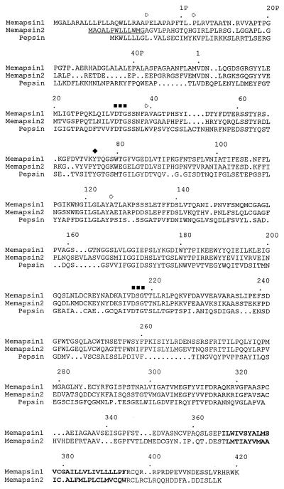
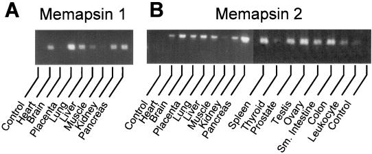
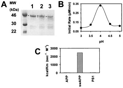
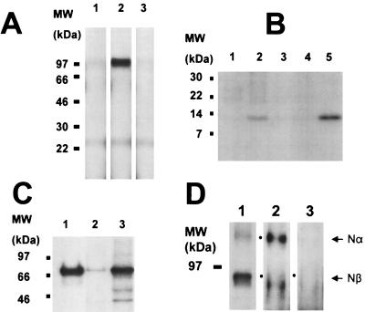
The cDNAs of two new human membrane-associated aspartic proteases, memapsin 1 and memapsin 2, have been cloned and sequenced. The deduced amino acid sequences show that each contains the typical pre, pro, and aspartic protease regions, but each also has a C-terminal extension of over 80 residues, which includes a single transmembrane domain and a C-terminal cytosolic domain. Memapsin 2 mRNA is abundant in human brain. The protease domain of memapsin 2 cDNA was expressed in Escherichia coli and was purified. Recombinant memapsin 2 specifically hydrolyzed peptides derived from the beta-secretase site of both the wild-type and Swedish mutant beta-amyloid precursor protein (APP) with over 60-fold increase of catalytic efficiency for the latter. Expression of APP and memapsin 2 in HeLa cells showed that memapsin 2 cleaved the beta-secretase site of APP intracellularly. These and other results suggest that memapsin 2 fits all of the criteria of beta-secretase, which catalyzes the rate-limiting step of the in vivo production of the beta-amyloid (Abeta) peptide leading to the progression of Alzheimer's disease. Recombinant memapsin 2 also cleaved a peptide derived from the processing site of presenilin 1, albeit with poor kinetic efficiency. Alignment of cleavage site sequences of peptides indicates that the specificity of memapsin 2 resides mainly at the S(1)' subsite, which prefers small side chains such as Ala, Ser, and Asp.


Figures

Figure 1
Figure 1
Predicted amino acid sequence of human prepro-memapsin 1 (GenBank accession no. AF200192) and prepro-memapsin 2 (GenBank accession no. AF200193) aligned against the sequence of human pre-pepsinogen. Amino acid codes are according to standard International Union of Pure and Applied Chemistry nomenclature. The beginning of pro and mature protease regions of pepsinogen are marked by residue numbers 1P and 1, respectively. Two active-site aspartic acids in D(T/S)G motifs are marked by ■ and conserved Tyr75 by ♦. Residues in the predicted transmembrane domains are in boldface.
Figure 2
Figure 2
Tissue distribution of M1 (A) and M2 (B) mRNA determined by reverse transcription–PCR visualized in agarose gel electrophoresis. Control experiment contained no template DNA. Amplification of glyceraldehyde-3-phosphate dehydrogenase mRNA produced uniform intensity (data not shown).
Figure 3
Figure 3
(A) Conversion of pro-M2pd (lane 1) at pH 4.0 to smaller fragments (lane 3) as shown by SDS/polyacrylamide electrophoretic pattern. Difference in migration between pro-M2pd and converted enzyme is evident in a mixture of the two (lane 2). Squares mark band positions. (B) Initial rate of hydrolysis of synthetic peptide swAPP (see Table 1) by M2pd at different pH. (C) Relative kcat/Km values for steady-state kinetic of hydrolysis of peptide substrates by M2pd (see text for peptides and values).
Figure 4
Figure 4
Processing of APP by M2 in HeLa cells. (A) SDS/PAGE patterns of immunoprecipitated APP Nβ-fragment (97-kDa band) from the condition media (2 h) of pulse–chase experiments. Lanes: 1, transfection of APP alone; 2, co-transfection of APP and M2; 3, same as lane 2 except that bafilomycin A1 is included. (B) SDS/PAGE patterns of APP βC-fragment (12 kDa) immunoprecipitated from the conditioned media of the same experiment as in A. Lanes: 1, transfection of APP; 2, co-transfection of APP and M2; 3, as in lane 2 but with bafilomycin A1; 4, transfection of Swedish APP; 5, co-transfection of Swedish APP and M2. (C) SDS/PAGE patterns of immunoprecipitated M2 (70 kDa). Lanes: 1, M2 transfected cells; 2, untransfected HeLa cells after long time film exposure; 3, endogenous M2 from HEK 293 cells. (D) SDS/PAGE patterns of APP fragments (100-kDa Nα-fragment and 95-kDa Nβ-fragment) recovered from conditioned media after immunoprecipitation using antibodies specific for the N-terminal region of APP. Lanes: 1, autoradiogram of immunoprecipitation from co-transfection of APP and M2; 2 and 3, immunoblotted by using antibody Aβ1–17, specific for the Nα-fragment that does not recognize the Nβ-fragment; 2, transfection with APP alone; 3, co-transfection of APP and M2. Dots mark band positions.
Figure 5
Figure 5
Intracellular localization of M2 and APP in a single cell. HeLa cells co-expressing APP and M2 were stained with antibodies directed toward APP (A, green image) and M2 (B, red image) and were visualized simultaneously by CSLM (see Materials and Methods) using a 100× objective. Areas of colocalization appear in yellow in overlay of two images in C.

Similar articles

Cited by

References

    1. Selkoe D J. Nature (London) 1999;399A:23–31. - PubMed
    1. Wolfe M S, Xia W, Ostaszewski B L, Diehl T S, Kimberly W T, Selkoe D J. Nature (London) 1999;398:513–517. - PubMed
    1. Thinakaran G, Borchelt D R, Lee M K, Slunt H H, Spitzer L, Kim G, Ratovitsky T, Davenport F, Nordstedt C, Seeger M, et al. Neuron. 1996;17:181–190. - PubMed
    1. Podlisny M B, Citron M, Amarante P, Sherrington R, Xia W, Zhang J, Diehl T, Levesque G, Fraser P, Haass C, et al. Neurobiol Dis. 1997;3:325–337. - PubMed
    1. Sinha S, Lieberburg I. Proc Natl Acad Sci USA. 1999;96:11049–11053. - PMC - PubMed

Publication types

Associated data