. 2000 Feb 15;97(4):1536-41.
doi: 10.1073/pnas.97.4.1536.

A 9-nt segment of a cellular mRNA can function as an internal ribosome entry site (IRES) and when present in linked multiple copies greatly enhances IRES activity

Affiliations

A 9-nt segment of a cellular mRNA can function as an internal ribosome entry site (IRES) and when present in linked multiple copies greatly enhances IRES activity

S A Chappell et al. Proc Natl Acad Sci U S A. .

Abstract

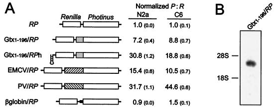
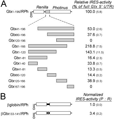
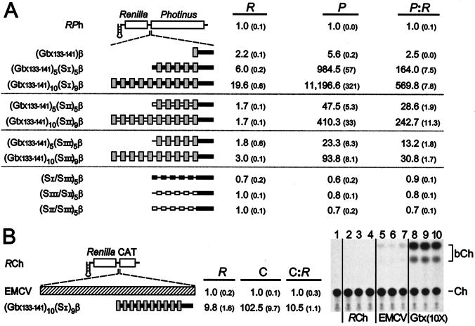
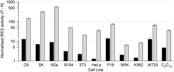
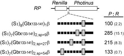
This study addresses the properties of a newly identified internal ribosome entry site (IRES) contained within the mRNA of the homeodomain protein Gtx. Sequential deletions of the 5' untranslated region (UTR) from either end did not define distinct IRES boundaries; when five nonoverlapping UTR fragments were tested, four had IRES activity. These observations are consistent with other cellular IRES analyses suggesting that some cellular IRESes are composed of segments (IRES modules) that independently and combinatorially contribute to overall IRES activity. We characterize a 9-nt IRES module from the Gtx 5' UTR that is 100% complementary to the 18S rRNA at nucleotides 1132-1124. In previous work, we demonstrated that this mRNA segment could be crosslinked to its complement within intact 40S subunits. Here we show that increasing the number of copies of this IRES module in the intercistronic region of a dicistronic mRNA strongly enhances IRES activity in various cell lines. Ten linked copies increased IRES activity up to 570-fold in Neuro 2a cells. This level of IRES activity is up to 63-fold greater than that obtained by using the well characterized encephalomyocarditis virus IRES when tested in the same assay system. When the number of nucleotides between two of the 9-nt Gtx IRES modules was increased, the synergy between them decreased. In light of these findings, we discuss possible mechanisms of ribosome recruitment by cellular mRNAs, address the proposed role of higher order RNA structures on cellular IRES activity, and suggest parallels between IRES modules and transcriptional enhancer elements.


Figures

Figure 1
Figure 1
Dicistronic analysis of Gtx IRES activity. (A) A schematic representation of the dicistronic constructs used in this analysis is indicated. Constructs are based on the RP (Renilla-Photinus) and RPh (Renilla-Photinus hairpin) vectors. Inserts include the full Gtx 5′ UTR (Gtx1–196), the EMCV and poliovirus IRESes, and the β-globin 5′ UTR. IRES activities are represented as ratios of Photinus luciferase activity to Renilla luciferase activity after transfection into N2a and C6 cells. Values represent activities that have been normalized to those of the RP vector. Numbers in parentheses represent SEM. (B) Northern blot of poly(A)+ RNA purified from N2a cells transfected with the Gtx1–196/RP dicistronic construct and probed with a fragment of the Photinus luciferase gene. The positions of the 28S and 18S rRNAs are indicated.
Figure 2
Figure 2
Deletion and fragment analysis of the Gtx 5′ UTR after transfection into N2a cells. (A) A schematic representation of the dicistronic constructs used in this analysis is indicated. Constructs are based on the RPh vector. Inserts include the full Gtx 5′ UTR (Gtx1–196) and deletions and fragments of this sequence. The Gtx construct designations indicate the exact nucleotide sequence present in each construct. IRES activities are represented as a percentage of the activity of the full Gtx 5′ UTR. Numbers in parentheses represent SEM. (B) Analysis of a 9-nt segment of the Gtx 5′ UTR. Construct β-globin/RPh contains the full 5′ UTR of the mouse β-globin mRNA. The activity of construct βGtx133–141/RPh is normalized to that of the β-globin control construct βglobin/RPh.
Figure 3
Figure 3
Dicistronic mRNAs containing synthetic IRESes based on multiple linked copies of nucleotides 133–141 of the Gtx 5′ UTR. (A) Schematic representation of the dicistronic constructs used in this analysis is indicated. Constructs are based on the RPh vector and contain 25 nt of the mouse β-globin 5′ UTR sequence (β) immediately upstream of the initiation codon, represented as a thick black line. (Inset) One, five, or ten copies of the Gtx IRES module (Gtx133–141) are indicated as gray bars; constructs with multiple copies are spaced 9 nt apart by one of three spacer sequences, SI, SII, or SIII, shown as a thick black bar, an open bar, and a thin line, respectively. Constructs were transfected into N2a cells, and luciferase activities were normalized for transfection efficiency (see Materials and Methods). Numbers in parentheses represent SEM. (B) Analysis of dicistronic constructs containing the CAT gene as the second cistron. The constructs used in this assay are based on the RCh vector. Inserts include the EMCV IRES and (Gtx133–141)10(SI)9β. Constructs were transfected into N2a cells and assayed for Renilla (R) and CAT (C) activities. CAT activities in the table were determined by liquid scintillation assay and, because CAT activity of the RCh vector was indistinguishable from a negative control, numbers were normalized to the activity of EMCV/RCh. An autoradiogram of a TLC assay of CAT activity is also shown. Lane 1, negative control; lanes 2–4, transfections with RCh; lanes 5–7, transfections with EMCV/RCh (EMCV); lanes 8–10, transfections with [Gtx133–141)10(SI)9β/RCh (Gtx(10X)]. Each lane represents an independent transfection. Ch represents 14C chloramphenicol; bCh represents butyrylated 14C chloramphenicol and is the product of CAT enzyme activity.
Figure 4
Figure 4
IRES activities in various cell lines. EMCV/RPh and (Gtx133–141)10(SI)9β/RPh were transfected into the cell lines indicated and IRES activity calculated as the ratio of Photinus to Renilla luciferase activities. These activities were normalized to those obtained with the RPh vector in these same cell lines. Activity of the EMCV/RPh construct is indicated as a black bar, and that of the (Gtx133–141)10(SI)9β/RPh construct is indicated as a gray bar. Vertical lines indicate SEM. Note that IRES activities are represented on a logarithmic scale. The following cell lines were used in this study: rat glial tumor line C6; human neuroblastoma SK-N-SH (SK); mouse neuroblastoma N2a; mouse fibroblast B104–1-1 (B104); mouse fibroblast NIH 3T3 (3T3); human epitheloid carcinoma HeLa; mouse embryonal carcinoma P19; rat normal kidney NRK; human chronic myelogenous leukemia K562; mouse pituitary tumor AtT-20 (AtT20); mouse muscle myoblast C2C12.
Figure 5
Figure 5
Effect on IRES activity of varying the spacing between two copies of the 9-nt Gtx IRES module (Gtx133–141). Constructs containing one or two copies of the 9-nt IRES module are indicated schematically. Constructs contain 25 nt of the mouse β-globin 5′ UTR sequence (β) immediately upstream of the initiation codon. Spacing between and upstream of the 9-nt IRES modules was achieved by using a 9-nt spacer sequence (SI). Spacing between two of the 9-nt IRES modules was 9, 27, or 54 nt. IRES activity is the ratio of Photinus to Renilla luciferase activity and is normalized to 100 for the activity of construct (SI)8(Gtx133–141)1β/RP. Numbers in parentheses represent SEM.

References

    1. Kozak M. Gene. 1999;234:187–208. - PubMed
    1. Gray N K, Wickens M. Annu Rev Cell Biol. 1998;14:399–458. - PubMed
    1. Jackson R J. In: Translational Control. Hershey J W B, Mathews M B, Sonenberg N, editors. Plainview, NY: Cold Spring Harbor Lab. Press; 1996. pp. 71–112.
    1. van der Velden A W, Thomas A A M. Int J Biochem Cell Biol. 1999;31:87–106. - PubMed
    1. Gingras A C, Raught B, Sonenberg N. Annu Rev Biochem. 1999;68:913–963. - PubMed

Publication types