. 2000 Feb 15;97(4):1713-8.
doi: 10.1073/pnas.97.4.1713.

Role of Syk in B-cell development and antigen-receptor signaling

Affiliations

Role of Syk in B-cell development and antigen-receptor signaling

R J Cornall et al. Proc Natl Acad Sci U S A. .

Abstract

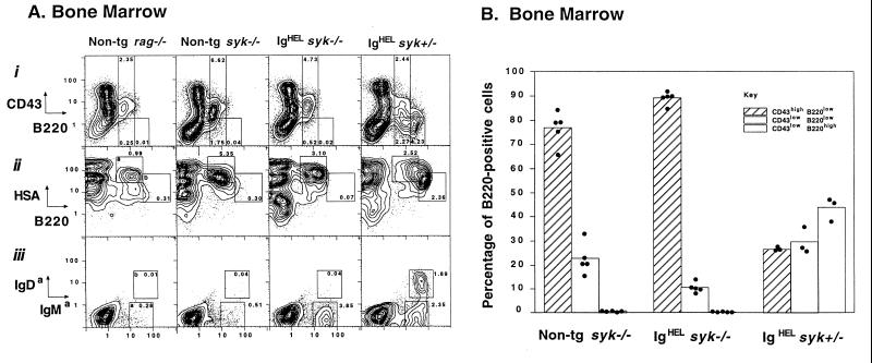
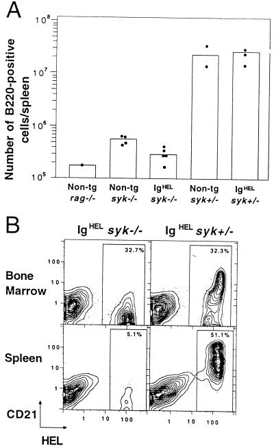
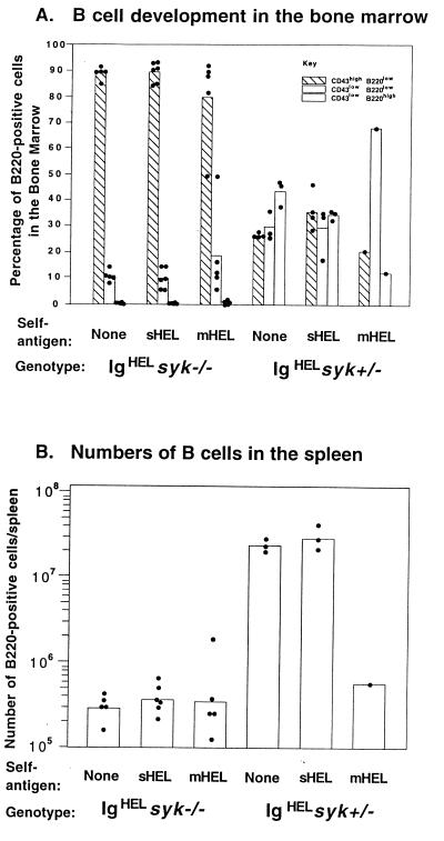
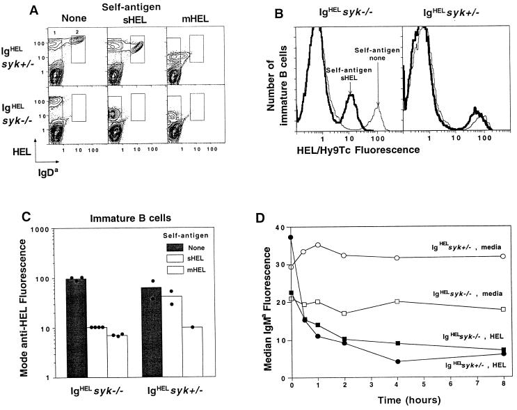
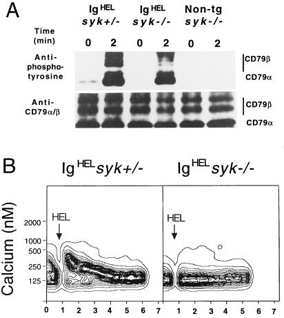
Antigen receptors (BCRs) on developing B lymphocytes play two opposing roles-promoting survival of cells that may later bind a foreign antigen and inhibiting survival of cells that bind too strongly to self-antigens. It is not known how these opposing outcomes are signaled by BCRs on immature B cells. Here we analyze the effect of a null mutation in the Syk tyrosine kinase on maturing B cells displaying a transgene-encoded BCR that binds hen egg lysozyme (HEL). In the absence of HEL antigen, HEL-specific BCRs are expressed normally on the surface of Syk-deficient immature B-lineage cells, but this fails to promote maturation beyond the earliest stages of B-lineage commitment. Binding of HEL antigen, nevertheless, triggers phosphorylation of CD79alpha/beta BCR subunits and modulation of receptors from the surface in Syk-deficient cells, but it cannot induce an intracellular calcium response. Continuous binding of low- or high-avidity forms of HEL, expressed as self-antigens, fails to restore the signal needed for maturation. Compared with the effects in the same system of null mutations in other BCR signaling elements, such as CD45 and Lyn kinase, these results indicate that Syk is essential for transmitting a signal that initiates the program of B-lymphocyte maturation.


Figures

Figure 1
Figure 1
Developmental arrest of Syk-deficient B cells in the bone marrow is not rescued by expression of rearranged Ig H + L transgenes encoding IgHEL. (A) B-cell development in bone marrow of nontransgenic rag−/−, nontransgenic syk−/−, and IgHEL-transgenic syk−/− and syk+/− chimeric mice, measured by two-color flow cytometry of cells stained for the indicated developmental markers. Windows in plots (iiiii) are conventional for immature (a) and mature (b) B cells, and the frequency of cells are shown as a percentage of the total cells in the bone marrow. Plots of syk−/− and syk+/− bone marrows are each representative of six chimeras. (B) Histograms summarizing the percentage of B220-positive B-lineage cells in the bone marrow which are CD43hiB220lo (Hardy A–C); CD43loB220lo (Hardy D–E); and CD43loB220hi (Hardy F). Bars represent arithmetic means and dots the percentages from individual chimeras. Mean numbers of B220 cells present in the bone marrow from one femur and one tibia of nontransgenic syk−/−, IgHEL syk−/− and IgHEL syk+/− mice were 20.10 × 10⁴ (SD = 7.60); 14.42 × 10⁴ (SD = 1.79); and 17.50 × 10⁴ (SD = 3.90), respectively (SD = standard deviation × 10−4).
Figure 2
Figure 2
The absence of syk−/− B cells in the blood and spleen is not corrected by expression of IgHEL. (A) Numbers of B220-positive cells in the spleen. Histograms show arithmetic means and dots represent individual mice which, with the exception of the rag−/− mouse, were all bone marrow chimeras. (B) Numbers and phenotype of B cells in the bone marrow and spleens of IgHEL-transgenic syk−/− and syk+/− chimeric mice, measured by two-color flow cytometry of cells stained for CD21 and HEL binding. Windows in plots show IgHEL transgenic B cells; the frequency of cells is shown as the percentage of the lymphocyte gate. Plots are each representative of six chimeras.
Figure 3
Figure 3
Engagement of the BCR by self-antigens cannot rescue the development of Syk-deficient B cells. (A) Development of syk+/− and syk−/− IgHEL B cells in the absence of autoantigen (none) or the presence of sHEL or mHEL. Data were obtained and presented as in Fig. 1. The result from the single IgHEL syk+/− mHEL chimera is representative of published data (21, 22). (B). Numbers of B220-positive syk+/− and syk−/− IgHEL cells in the absence of autoantigen or the presence of sHEL or mHEL. Histograms show arithmetic means and dots represent individual chimeras.
Figure 4
Figure 4
Modulation of IgM in response to antigen does not depend on Syk. (A) Two-color flow cytometry of bone marrow cells from syk+/− and syk−/− IgHEL chimeric mice in the absence of autoantigen or the presence of sHEL or mHEL. The windows containing immature (1) and mature (2) B cells are indicated. Plots are representative of six chimeras. (B) Overlaid histograms of IgM expression (HEL/Hy9Tc fluorescence) on immature IgHEL B cells in the presence or absence of sHEL autoantigen. (C) Mode expression of HEL-binding BCRs on the surface of immature IgHEL B cells from syk−/− and syk+/− chimeric mice, developing in the absence of autoantigen or the presence of sHEL or mHEL. Histograms represent arithmetic means and dots individual animals. (D) Median IgMa receptor levels on naive, immature B220lo IgHEL B cells from the bone marrow of syk−/− (squares) or syk+/− (circles) mice, after incubating in vitro for varying lengths of time with 1 μg/ml HEL (closed points) or with media alone (open points).
Figure 5
Figure 5
Syk is not required for antigen-induced phosphorylation of CD79α and CD79β but is essential for a calcium response. (A) Western blot of immunoprecitated CD79β from lysates of syk−/− and syk+/− IgHEL and syk−/− nontransgenic bone marrow cells. Cells were stimulated with HEL (1 μg/ml) for 2 min or left unstimulated. Panels show induced tyrosine phosphorylation by probing the immunoprecipitates for antiphosphotyrosine (4G10), relative to the total immunoprecipitated protein measured by reprobing the same blot with antisera to CD79α and β. Results are representative of three experiments. (B) Flow cytometry of indo-1-loaded cells was used to monitor intracellular calcium levels as a function of time after stimulation of IgHEL syk−/− and syk+/− B cells from bone marrow. All measurements were gated on immature, B220lo cells. Arrows indicate time of addition of HEL antigen (1 μg/ml). Equivalent results were obtained from three separate experiments.

References

    1. Healy J I, Goodnow C C. Annu Rev Immunol. 1998;16:645–670. - PubMed
    1. Rolink A, Melchers F. Adv Immunol. 1993;53:123–156. - PubMed
    1. Rajewsky K. Nature (London) 1996;381:751–758. - PubMed
    1. Papavasiliou F, Misulovin Z, Suh H, Nussenzweig M C. Science. 1995;268:408–411. - PubMed
    1. Papavasiliou F, Janokovic M, Suh H, Nussenzweig M C. J Exp Med. 1995;182:1389–1394. - PMC - PubMed

Publication types

MeSH terms