. 2000 Feb 15;97(4):1766-71.
doi: 10.1073/pnas.040545897.

Identification of a Plasmodium falciparum intercellular adhesion molecule-1 binding domain: a parasite adhesion trait implicated in cerebral malaria

Affiliations

Identification of a Plasmodium falciparum intercellular adhesion molecule-1 binding domain: a parasite adhesion trait implicated in cerebral malaria

J D Smith et al. Proc Natl Acad Sci U S A. .

Erratum in

  • Proc Natl Acad Sci U S A. 2006 Aug 8;103(32):12209. Fagen, T [corrected to Fagan, T]

Abstract

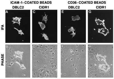
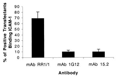
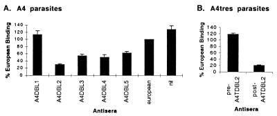
Binding of infected erythrocytes to brain venules is a central pathogenic event in the lethal malaria disease complication, cerebral malaria. The only parasite adhesion trait linked to cerebral sequestration is binding to intercellular adhesion molecule-1 (ICAM-1). In this report, we show that Plasmodium falciparum erythrocyte membrane protein 1 (PfEMP1) binds ICAM-1. We have cloned and expressed PfEMP1 recombinant proteins from the A4tres parasite. Using heterologous expression in mammalian cells, the minimal ICAM-1 binding domain was a complex domain consisting of the second Duffy binding-like (DBL) domain and the C2 domain. Constructs that contained either domain alone did not bind ICAM-1. Based on phylogenetic criteria, there are five distinct PfEMP1 DBL types designated alpha, beta, gamma, delta, and epsilon. The DBL domain from the A4tres that binds ICAM-1 is DBLbeta type. A PfEMP1 cloned from a distinct ICAM-1 binding variant, the A4 parasite, contains a DBLbeta domain and a C2 domain in tandem arrangement similar to the A4tres PfEMP1. Anti-PfEMP1 antisera implicate the DBLbeta domain from A4var PfEMP1 in ICAM-1 adhesion. The identification of a P. falciparum ICAM-1 binding domain may clarify mechanisms responsible for the pathogenesis of cerebral malaria and lead to interventions or vaccines that reduce malarial disease.


Figures

Figure 1
Figure 1
Schematic of A4tres and A4var PfEMP1 domain organization and expression constructs. The entire extracellular region of PfEMP1, including a putative transmembrane domain, is encoded in exon 1. The intron and exon 2 (partially sequenced for each PfEMP1) are labeled. Recombinant PfEMP1 proteins expressed in Cos-7 cells are shown beneath the protein schematics with domain boundaries listed. The A4var DBLβC2 was synthesized and tested for ICAM-1 binding in this paper. Previously, other A4var PfEMP1 recombinant proteins that covered the whole extracellular domain were tested for ICAM-1 binding (21).
Figure 2
Figure 2
A4tres PfEMP1 binding domains for ICAM-1 and CD36. ICAM-1-coated beads specifically bind DBLβC2-transfected cells (A and B) and do not bind CIDR1 transfectants (C and D). In contrast, CD36-coated beads bind CIDR1-transfected cells (G and H) but do not adhere to DBLβC2 transfectants (E and F). (Top; A, C, E, and G) Immunofluorescent images from the staining of transfected cells with an antibody against an epitope tag in the recombinant protein. (Bottom; B, D, F, and H) Phase images.
Figure 3
Figure 3
Binding of PfEMP1 recombinant proteins to ICAM-1 and CD36. PfEMP1 recombinant proteins were expressed at the surface of Cos-7 cells and tested for binding to ICAM-1/Fc or CD36-coated dynal magnetic beads. The percentage of binding for each PfEMP1 recombinant proteins was calculated by counting 100 positive transfectants and scoring Cos-7 cells with 5 or more beads as positive. (A and B) Binding to ICAM-1/Fc. (C) Binding to CD36. Results in B and C are the mean of binding plus standard deviation of experiments performed in duplicate.
Figure 4
Figure 4
Reversal of A4tres DBLβC2 recombinant protein binding to ICAM-1 with ICAM-1 antibodies. A4tres DBLβC2-transfected Cos-7 cells were incubated with ICAM-1/Fc coated beads, washed by inversion, and then incubated in the presence or absence of 10 μg/ml domain 1-specific ICAM-1 antibodies (1G12, 15.2, or RR1/1) or normal mouse IgG. Binding was quantified by inspecting 100 positive transfectants and scoring Cos-7 cells with 5 or more beads as positive. The percentage binding has been normalized to binding observed in the presence of normal mouse IgG. Results are the mean of six experiments plus standard error.
Figure 5
Figure 5
Alignments of A4tres and A4var PfEMP1 DBLβC2 domains. The start of DBLβ and C2 domains is labeled above the alignment. Dashes (-) indicate gaps introduced to maintain alignment. A 100% amino acid consensus for eight DBLβC2 domains, including A4tres and A4var, is shown below the multiple alignment. Invariant residues are identified in bold with capital letters of the single amino acid code. Residues with a similar amino acid character are shown in bold: c (charge), h (hydrophobic), p (polar), s (small), u (tiny), b (big). Shown below the consensus is a secondary structural prediction of folding for the A4tres DBLβC2. H stands for α helical; β strands were not predicted. The structural prediction is highly representative of the seven other sequences in the alignment; helical boundaries were identical or varied by only a few amino acids (data not shown).
Figure 6
Figure 6
Reversal of A4 or A4tres parasite binding to ICAM-1 with PfEMP1 antisera. Infected erythrocytes were bound to ICAM-1 coated onto plastic, and the binding was reversed by using PfEMP1 antisera raised against different DBL domains. (A) A4-infected erythrocyte treated with A4var PfEMP1 antisera, nonimmune European sera, or no antibody (nil). (B) A4tres-infected erythrocytes treated with preimmune sera or anti-DBLβ sera (postimmune). (A and B) The binding is presented as a percentage of that observed using nonimmune European sera for reversal. Results in A are the mean of four experiments and in B the mean of two experiments plus standard error.

References

    1. Warrell D A, Molyneux M E, Beales P F. Trans R Soc Trop Med Hyg. 1990;84:1–65.
    1. Miller L H, Good M F, Milon G. Science. 1994;264:1878–1883. - PubMed
    1. MacPherson G G, Warrell M J, White N J, Looareesuwan S, Warrell D A. Am J Pathol. 1985;119:385–401. - PMC - PubMed
    1. Pongponratn E, Riganti M, Punpoowong B, Aikawa M. Am J Trop Med Hyg. 1991;44:168–175. - PubMed
    1. Barnwell J W, Asch A S, Nachman R L, Yamaya M, Aikawa M, Ingravallo P. J Clin Invest. 1989;84:765–772. - PMC - PubMed

Publication types

MeSH terms