. 2000 Feb 15;97(4):1879-84.
doi: 10.1073/pnas.97.4.1879.

Dopamine tone regulates D1 receptor trafficking and delivery in striatal neurons in dopamine transporter-deficient mice

Affiliations

Dopamine tone regulates D1 receptor trafficking and delivery in striatal neurons in dopamine transporter-deficient mice

B Dumartin et al. Proc Natl Acad Sci U S A. .

Abstract

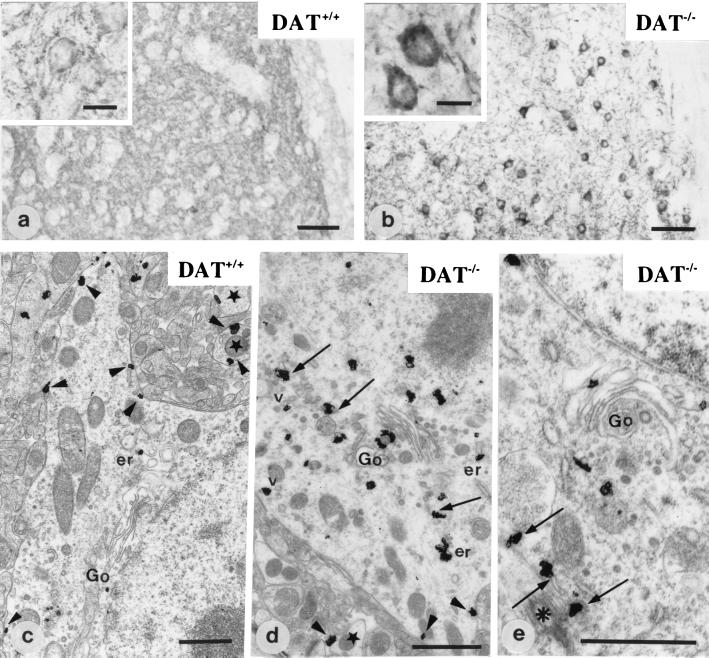
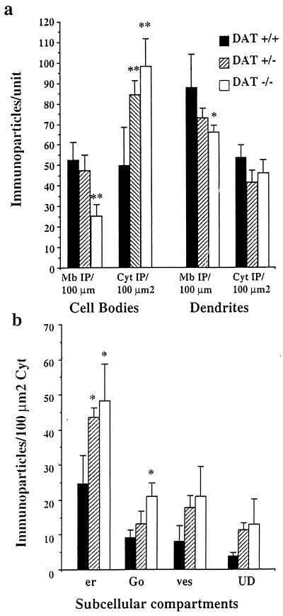
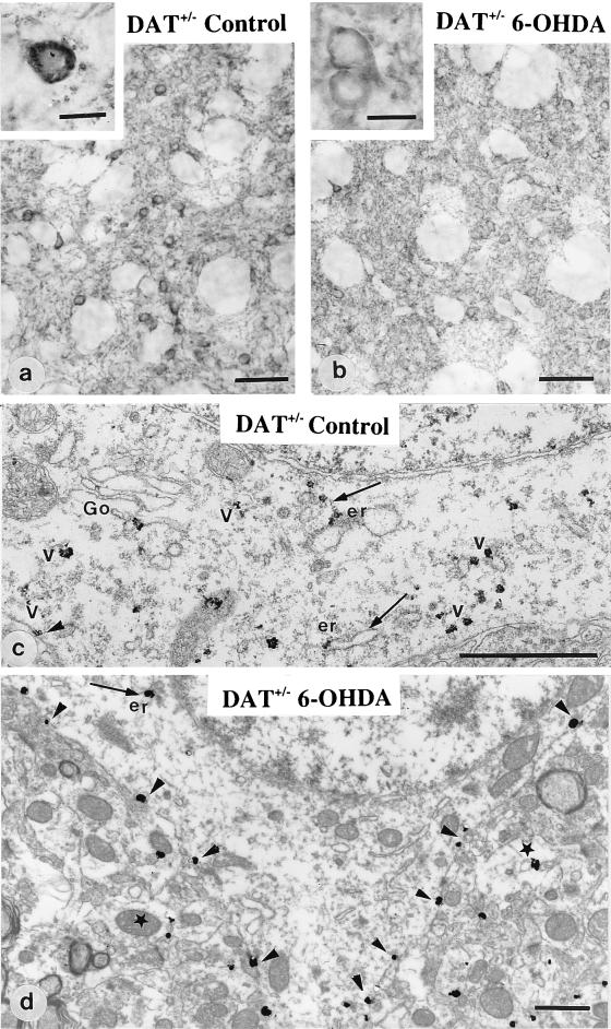
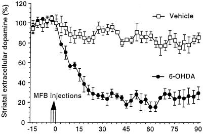
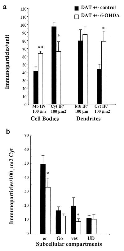
In vivo, G protein-coupled receptors (GPCR) for neurotransmitters undergo complex intracellular trafficking that contribute to regulate their abundance at the cell surface. Here, we report a previously undescribed alteration in the subcellular localization of D1 dopamine receptor (D1R) that occurs in vivo in striatal dopaminoceptive neurons in response to chronic and constitutive hyperdopaminergia. Indeed, in mice lacking the dopamine transporter, D1R is in abnormally low abundance at the plasma membrane of cell bodies and dendrites and is largely accumulated in rough endoplasmic reticulum and Golgi apparatus. Decrease of striatal extracellular dopamine concentration with 6-hydroxydopamine (6- OHDA) in heterozygous mice restores delivery of the receptor from the cytoplasm to the plasma membrane in cell bodies. These results demonstrate that, in vivo, in the central nervous system, the storage in cytoplasmic compartments involved in synthesis and the membrane delivery contribute to regulate GPCR availability and abundance at the surface of the neurons under control of the neurotransmitter tone. Such regulation may contribute to modulate receptivity of neurons to their endogenous ligands and related exogenous drugs.


Figures

Figure 1
Figure 1
Immunohistochemical detection of D1R at the light and electron microscopic level in DAT+/+ (a and c) and DAT−/− mice (b, d, and e). In wild-type mice (DAT+/+) (a and c), D1R is located in the neuropile and at the plasma membrane of cell bodies (a, Inset; and c, arrowheads). Stars point to dendrite profiles. Ultrastructural localization of D1R in c shows very few intracytoplasmic immunoparticles associated with the Golgi apparatus (Go) and the endoplasmic reticulum (er). In DAT−/− mice (b, d, and e), D1R is located primarily in the cytoplasm of cell bodies (b, Inset). The receptor is associated with the Golgi apparatus (Go), the endoplasmic reticulum (arrows and er), and some vesicles (v). A few immunoparticles are detected along the plasma membrane (d, arrowheads). In e, part of D1R immunoreactivity associated with endoplasmic reticulum (arrows) is in the pericentriolar area (asterisk). [Scale bar = 50 μm (a and b), 10 μm (a and b, Insets), and 1 μm (c, d, and e)].
Figure 2
Figure 2
Quantitative analysis of the subcellular distribution of D1R at the electron microscopic level. (a) Measure of D1R immunoreactivity at the plasma membrane and in the cytoplasm: The number of immunoparticles +/− SEM was counted in relation to the plasma membrane length (Mb IP/100 μm) and to the cytoplasmic surface (Cyt IP/100 μm2) in cell bodies and dendrites. DAT−/− neurons show a sharp and significant decrease of D1R at the plasma membrane of cell bodies and dendrites as compared with DAT+/+ neurons. They also display sharp increase of intracytoplasmic immunoparticles in the cell bodies. DAT+/− mice display significant increase of D1R in the cytoplasm without significant reduction at the plasma membrane. (b) Measure of D1R immunoreactivity in the cytoplasmic organelles in the cell bodies. In DAT+/+ cell bodies, the largest part of cytoplasmic D1R is detected in association with the endoplasmic reticulum (er) and the Golgi apparatus (Go). Other particles are present in vesicles (ves) or in an unidentified cytoplasmic compartment (UD). Immunoparticles density is dramatically increased in the endoplasmic reticulum and in the Golgi apparatus in DAT−/− mice and only in the endoplasmic reticulum in DAT+/− mice. **, P ≤ 0.01; *, P < 0.05.
Figure 3
Figure 3
Immunohistochemical detection of D1R at the light and electron microscopic level in heterozygous mice unilaterally treated with 6–0HDA. In the control side (a and c), striatal neurons display D1R mostly located in the cytoplasm, associated with the Golgi apparatus (Go), the endoplasmic reticulum (er), and vesicles (arrows). In the 6-OHDA-injected side (b and d), D1R appears homogeneously distributed in the neuropile and largely redistributed at the plasma membrane of cell bodies (b, Inset; and d, arrowheads) with a limited number of immunoparticles in the cytoplasm. Star points to a dendritic profile. (Magnification bar: a and b = 50 μm; a and b, Insets = 10 μm; c and d = 1 μm).
Figure 4
Figure 4
Effect of 6-OHDA injection in the MFB on the extracellular dopamine concentration in the striatum of DAT+/− mice. The extracellular dopamine concentration was monitored every 90 s in the striatum with an electrochemically treated carbon fiber electrode combined with differential pulse voltammetry. Injections of the 6-OHDA solution or of the vehicle were performed after a control period of 15 min. The extracellular dopamine level was expressed in percent of the mean of the 10 absolute peak amplitudes recorded before injections. 6–0HDA provokes a rapid and dramatic decrease of the dopamine content as compared with control experiment with vehicle.
Figure 5
Figure 5
Quantitative analysis of the subcellular distribution of D1R in DAT+/− mice after 6-OHDA injection. (a) Measure of D1R immunoreactivity at the plasma membrane and in the cytoplasm: The number of immunoparticles +/− SEM was counted in relation to the plasma membrane length (Mb IP/100 μm) and to the cytoplasmic surface (Cyt IP/100 μm2) in cell bodies and dendrites. Cell bodies in 6-OHDA-injected side display decreased cytoplasmic D1R and increased D1R at the plasma membrane. Dendrites display unchanged membrane-bound receptor but increased cytoplasmic D1R. (b) Measure of D1R immunoreactivity in the cytoplasmic organelles in the cell bodies: the endoplasmic reticulum (er) and the vesicles (ves) display a significant decrease of D1R in 6-OHDA-injected side as compared with the control side. **, P ≤ 0.001; *, P ≤ 0.05.

References

    1. Hoyer D, Humphrey P P. J Recept Signal Transduct Res. 1997;17:551–568. - PubMed
    1. Sokoloff P, Schwartz J C. Trends Pharmacol Sci. 1995;16:270–274. - PubMed
    1. Hersch S M, Ciliax B J, Guterkunst C A, Rees H D, Heilman C J, Yung K K, Bolam J P, Ince E, Yi H, Levey A I. J Neurosci. 1994;14:3351–3363.
    1. Liu H, Brown J L, Jasmin L, Maggio J E, Vigna S R, Mantyh P W, Basbaum A I. Proc Natl Acad Sci USA. 1994;91:1009–1013. - PMC - PubMed
    1. Levey A I, Edmunds S M, Hersch S M, Wiley R G, Heilman C J. J Comp Neurol. 1995;351:339–356. - PubMed

Publication types

MeSH terms

LinkOut - more resources