. 2000 Mar;68(3):1156-63.
doi: 10.1128/IAI.68.3.1156-1163.2000.

Synthetic peptide immunogens elicit polyclonal and monoclonal antibodies specific for linear epitopes in the D motifs of Staphylococcus aureus fibronectin-binding protein, which are composed of amino acids that are essential for fibronectin binding

Affiliations

Synthetic peptide immunogens elicit polyclonal and monoclonal antibodies specific for linear epitopes in the D motifs of Staphylococcus aureus fibronectin-binding protein, which are composed of amino acids that are essential for fibronectin binding

M Huesca et al. Infect Immun. 2000 Mar.

Abstract

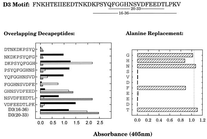
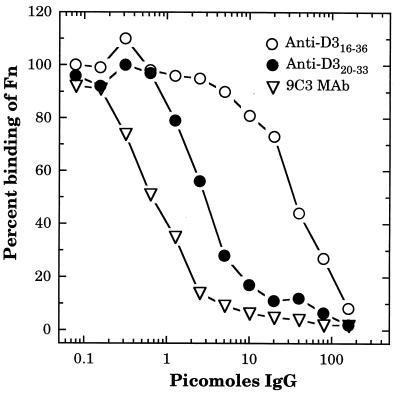
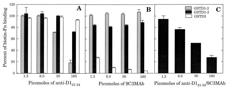
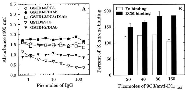
A fibronectin (Fn)-binding adhesin of Staphylococcus aureus contains three tandem 37- or 38-amino-acid motifs (D1, D2, and D3), which function to bind Fn. Plasma from patients with S. aureus infections contain antibodies that preferentially recognize ligand induced binding sites in the D motifs and do not inhibit Fn binding (F. Casolini, L. Visai, D. Joh, P. G. Conaldi, A. Toniolo, M. Höök, and P. Speziale, Infect. Immun. 66:5433-5442, 1998). To eliminate the influence of Fn binding on antibody development, we used synthetic peptide immunogens D1(21-34) and D3(20-33), which each contain a conserved pattern of amino acids that is essential for Fn binding but which cannot bind Fn without N- or C-terminal extensions. The D3(20-33) immunogen promoted the production of polyclonal antibodies that were 10-fold more effective as inhibitors of Fn-binding to the D3 motif than antibodies obtained by immunizing with an extended peptide D3(16-36), which exhibits functional Fn binding. The D3(20-33) immunogen also facilitated the production of a monoclonal antibody, 9C3, which was highly specific for the epitope SVDFEED, and abolished Fn binding by the D3 motif. When mixed with polyclonal anti-D1(21-34) immunoglobulin G, 70% inhibition of Fn binding to the three tandem D motifs was achieved compared to no more than 30% inhibition with either antibody preparation alone. Therefore, by immunizing with short synthetic peptides that are unable to bind Fn, we have effectively stimulated the production of antibodies specific for epitopes comprised of amino acids that are essential for Fn binding. Although these epitopes occur within a conserved pattern of amino acids that is required for Fn binding, the antibodies recognized specific linear epitope sequences and not a conserved structure common to all repeated motifs.


Figures

FIG. 1
FIG. 1
Epitope mapping of anti-D316–36 and anti-D320–33 antibodies. Biotin-labeled synthetic peptides (2 μg · ml−1) were captured on streptavidin-coated microtiter plates, followed by incubation with 1 μg of anti-D316–36 or anti-D320–33 polyclonal IgG per ml or 50 ng of monoclonal anti-D320–33 IgG (MAb 9C3) per ml. Plates were developed as in the standard ELISA protocol. The sequence of the intact D3 motif, with the underlined amino acids representing the D316–36 and D320–33 sequences is displayed at the top of the figure. The left panel displays the comparison of reactivity between the polyclonal anti-D316–36 (■) and anti-D320–33 (□) IgG and the MAb 9C3 (▨), toward a series of overlapping decapeptides spanning amino acids 11 to 37 of the D3 motif. The right panel displays the reactivity of MAb 9C3 toward a series of peptides containing a single alanine substitution at each of the indicated amino acids within the sequence D323–33. The dashed line represents the response to D323–33 with no alanine substitutions. Each value represents the average of triplicate determinations.
FIG. 2
FIG. 2
Recombinant Fn-binding peptides separated by SDS-PAGE and stained with Coomassie blue (A and D) or transferred to Immobilon-P membrane, followed by detection with 1 μg of biotin-labeled N29 fragment of Fn (B and E) or MAb 9C3 (C and F) per ml. Lanes (A, B, and C): 1, GST protein; 2, GSTD1-3; 3, GST-D1-2; 4, GSTD2-3; 5, GSTD3. Lanes (D, E, and F): 1, GST protein; 2, GSTA1-3, and 3, GSTB1-3 (both from S. dysgalactiae); 4, rFNBD-P (from S. pyogenes); 5, GSTD3. Protein loading was 1 μg per lane in panels A, D, E, and F, except for lane 5 in panels E and F, which contained 25 and 100 ng, respectively. In panels B and C, 25 and 100 ng of protein per lane, respectively, was used.
FIG. 3
FIG. 3
Inhibition of Fn binding to GSTD3 by anti-D316–36 and anti-D320–33 antibodies. GSTD3-coated microtiter plates were incubated for 60 min with 50 μl of various concentrations of anti-D316–36 or anti-D320–33 polyclonal IgG or anti-D320–33 monoclonal IgG (MAb 9C3), followed by the addition of 28 ng in 50 μl per well of biotinylated Fn. After incubation for an additional 60 min, bound Fn was detected with streptavidin-alkaline phosphatase conjugate. Absorbance values (A405) were determined after 60 min of development and then converted into percent values, with 100% representing the A405 value in the absence of added IgG. Each point represents the average from triplicate determinations.
FIG. 4
FIG. 4
Effect of specific antibodies on the binding of biotinylated Fn to different Fn-binding GST fusion proteins. Wells of microtiter plates were coated with 500 ng of GSTD3, GSTD1-2, or GSTD1-3 and then incubated for 60 min with 50 μl of various concentrations of anti-D121–34 polyclonal IgG (A), anti-D32033 MAb 9C3 IgG (B), or combined anti-D121–34 polyclonal IgG and MAb 9C3 (C), followed by incubation for an additional 60 min with 28 ng (in 50 μl) of biotinylated Fn per well. Bound Fn was detected with streptavidin-alkaline phosphatase conjugate. Each point represents the average from triplicate determinations.
FIG. 5
FIG. 5
Effect of specific antibodies on binding of biotin-labeled GST D-motif fusion proteins to the wells of microtiter plates coated with 500 ng of Fn per well (A) and on the binding of biotin-labeled S. aureus MRSA strain 34 to the wells of microtiter plates coated with 500 ng per well of Fn or ECM deposited by keratinocyte cell culture (B). In panel A, 1 μg of biotin-labeled fusion proteins was preincubated for 60 min with the indicated amount of polyclonal D121–34 IgG, MAb 9C3 IgG, or both combined in a volume of 100 μl prior to addition to Fn-coated microtiter plates. In panel B, 107 biotinylated S. aureus cells were preincubated for 60 min with a mixture of D121–34 IgG and MAb 9C3 prior to addition to microtiter plates coated with Fn or keratinocyte ECM. Binding was detected with streptavidin-alkaline phosphatase conjugate. Each point represents the average of duplicate determinations.

References

    1. Appel R D, Bairoch A, Hochstrasser D F. A new generation of information retrieval tools for biologists: the example of the ExPASy WWW server. Trends Biochem Sci. 1994;19:258–260. - PubMed
    1. Archer G L. Staphylococcus aureus: a well-armed pathogen. Clin Infect Dis. 1998;26:1179–1181. - PubMed
    1. Balaban N, Goldkorn T, Nhan R T, Dang L B, Scott S, Ridgley R M, Rasooly A, Wright S C, Larrick J W, Rasooly R, Carlson J R. Autoinducer of virulence as a target for vaccine and therapy against Staphylococcus aureus. Science. 1998;280:438–440. - PubMed
    1. Brennan F R, Jones T D, Longstaff M, Chapman S, Bellaby T, Smith H, Xu F, Hamilton W D, Flock J I. Immunogenicity of peptides derived from a fibronectin-binding protein of S. aureus expressed on two different plant viruses. Vaccine. 1999;17:1846–1857. - PubMed
    1. Casolini F, Visai L, Joh D, Conaldi P G, Toniolo A, Hook M, Speziale P. Antibody response to fibronectin-binding adhesin FnbpA in patients with Staphylococcus aureus infections. Infect Immun. 1998;66:5433–5442. - PMC - PubMed

Publication types

LinkOut - more resources