

doi: 10.1128/IAI.68.3.1710-1713.2000.
Intestinal epithelial cell apoptosis following Cryptosporidium parvum infection
Affiliations
- PMID: 10678994
- PMCID: PMC97335
- DOI: 10.1128/IAI.68.3.1710-1713.2000
Item in Clipboard
Intestinal epithelial cell apoptosis following Cryptosporidium parvum infection
Infect Immun.
2000 Mar.
Abstract
Cryptosporidium parvum induces moderate levels of apoptosis of cultured human intestinal epithelial cells, which are maximal at 24 h after infection. Apoptosis is further increased in C. parvum-infected cells by inhibition of NF-kappaB. C. parvum infection also attenuates epithelial apoptosis induced by strongly proapoptotic agents. The data suggest C. parvum has developed strategies to limit apoptosis in order to facilitate its growth and maturation in the early period after epithelial cell infection.
Figures
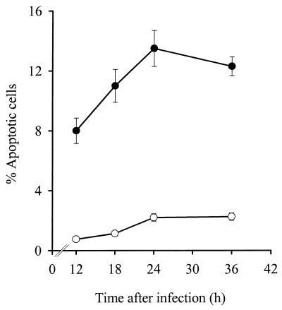
Time course of apoptosis in HCT-8 cells infected with C. parvum. Apoptotic cells were determined by staining with Hoechst 33258 dye. The percentage of apoptotic cells is shown for C. parvum-infected cells (●) and uninfected control cells (○). Data are means ± standard errors of four repeated experiments. Similar findings were obtained by assessing caspase cleavage of keratin 18 as a measure of apoptosis (data not shown).
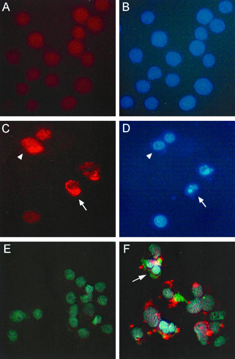
Colocalization of C. parvum infection and apoptosis in HCT-8 cells. Confluent monolayers of uninfected (A and B) and C. parvum-infected (C and D) HCT-8 cells were fixed 24 h after infection and stained for C. parvum and with Hoechst 33258 dye. Panels A and C are photomicrographs obtained with an Omega optical XF34 fluorescence filter to visualize C. parvum staining, while panels B and D are photomicrographs from the respective identical fields obtained with an Omega optical XF05 filter to visualize staining with Hoechst dye. A cell infected with C. parvum and undergoing apoptosis is indicated by the arrows, and a nonapoptotic cell infected with C. parvum is indicated by the solid arrowheads. Panels E and F display triple staining of uninfected (E) and C. parvum-infected (F) HCT-8 cultures. C. parvum was stained with Cy3-conjugated secondary antibody (red), nuclear morphology was stained with Hoechst 33258 dye (blue), and cleaved cytokeratin 18 was detected with a Cy2-conjugated secondary antibody (green), which was visualized with an Omega optical XF23 filter. Triple-staining images were layered by using the program Adobe Photoshop. The arrow depicts a C. parvum-infected cell undergoing apoptosis, as indicated by apoptotic nuclear morphology and the presence of cleaved cytokeratin 18. Overlapping areas of green and red appear as yellow. Original magnification, ×630.
References
-
- Argenzio R A, Lecce J, Powell D W. Prostanoids inhibit intestinal NaCl absorption in experimental porcine cryptosporidiosis. Gastroenterology. 1993;104:440–447. - PubMed
-
- Argenzio R A, Liacos J A, Levy M L, Meuten D J, Lecce J G, Powell D W. Villous atrophy, crypt hyperplasia, cellular infiltration, and impaired glucose-Na absorption in enteric cryptosporidiosis of pigs. Gastroenterology. 1990;98:1129–1140. - PubMed
-
- Beg A A, Baltimore D. An essential role for NF-κB in preventing TNF-α-induced cell death. Science. 1996;274:782–784. - PubMed
-
- Chen X-M, Gores G J, Paya C V, LaRusso N F. Cryptosporidium parvum induces apoptosis in biliary epithelia by a Fas/Fas ligand-dependent mechanism. Am J Physiol. 1999;277:G599–G608. - PubMed
Publication types
MeSH terms
Substances
Grants and funding
LinkOut - more resources
Full Text Sources
