. 2000 Feb;11(2):467-80.
doi: 10.1091/mbc.11.2.467.

Rab7: a key to lysosome biogenesis

Affiliations
Free PMC article

Rab7: a key to lysosome biogenesis

C Bucci et al. Mol Biol Cell. 2000 Feb.
Free PMC article

Abstract

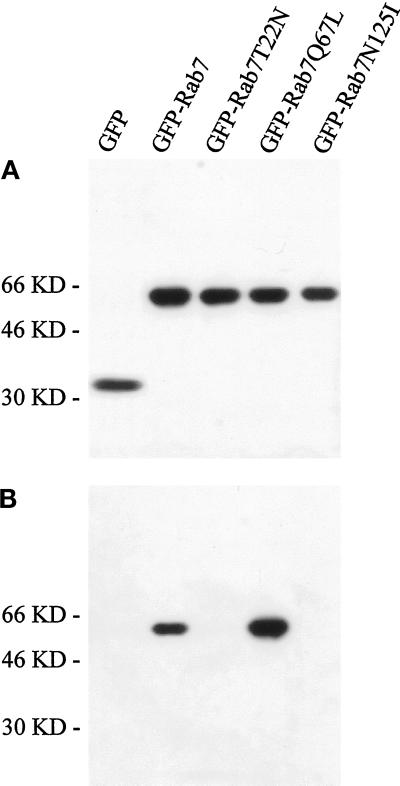
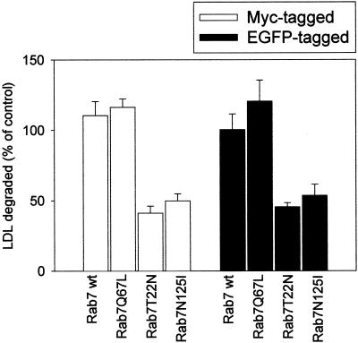
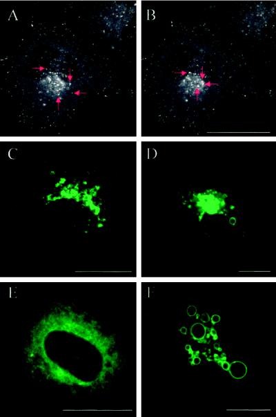
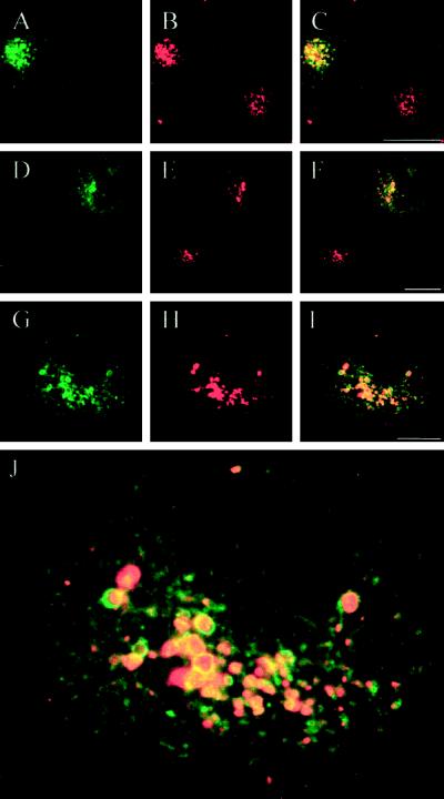
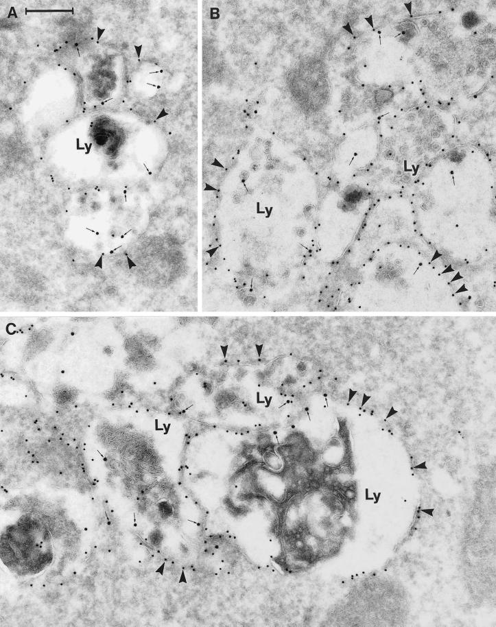
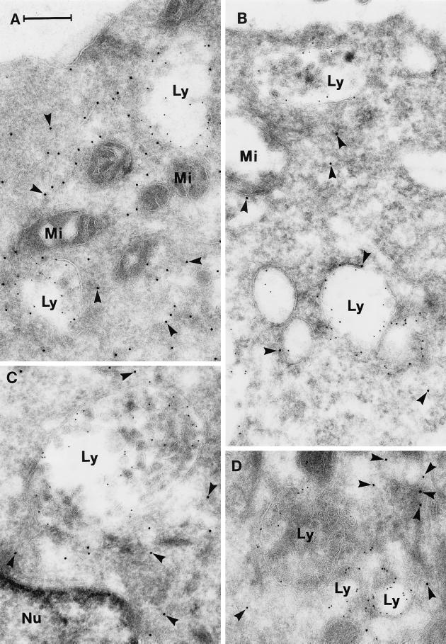
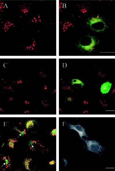
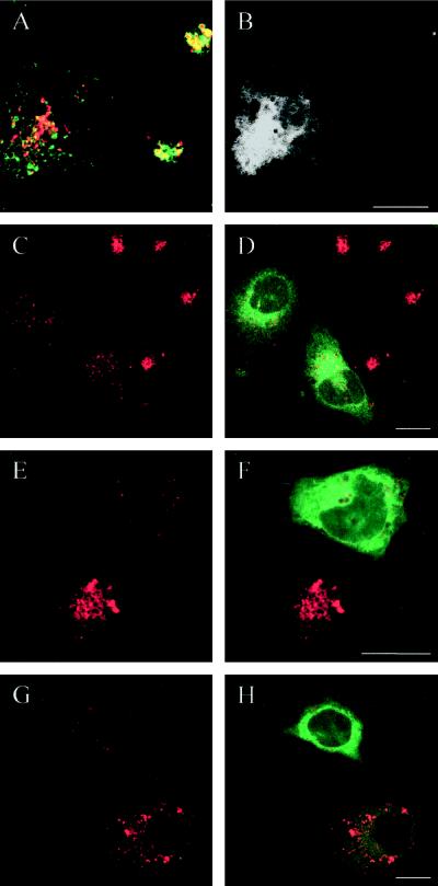
The molecular machinery behind lysosome biogenesis and the maintenance of the perinuclear aggregate of late endocytic structures is not well understood. A likely candidate for being part of this machinery is the small GTPase Rab7, but it is unclear whether this protein is associated with lysosomes or plays any role in the regulation of the perinuclear lysosome compartment. Previously, Rab7 has mainly been implicated in transport from early to late endosomes. We have now used a new approach to analyze the role of Rab7: transient expression of Enhanced Green Fluorescent Protein (EGFP)-tagged Rab7 wt and mutant proteins in HeLa cells. EGFP-Rab7 wt was associated with late endocytic structures, mainly lysosomes, which aggregated and fused in the perinuclear region. The size of the individual lysosomes as well as the degree of perinuclear aggregation increased with the expression levels of EGFP-Rab7 wt and, more dramatically, the active EGFP-Rab7Q67L mutant. In contrast, upon expression of the dominant-negative mutants EGFP-Rab7T22N and EGFP-Rab7N125I, which localized mainly to the cytosol, the perinuclear lysosome aggregate disappeared and lysosomes, identified by colocalization of cathepsin D and lysosome-associated membrane protein-1, became dispersed throughout the cytoplasm, they were inaccessible to endocytosed molecules such as low-density lipoprotein, and their acidity was strongly reduced, as determined by decreased accumulation of the acidotropic probe LysoTracker Red. In contrast, early endosomes associated with Rab5 and the transferrin receptor, late endosomes enriched in the cation-independent mannose 6-phosphate receptor, and the trans-Golgi network, identified by its enrichment in TGN-38, were unchanged. These data demonstrate for the first time that Rab7, controlling aggregation and fusion of late endocytic structures/lysosomes, is essential for maintenance of the perinuclear lysosome compartment.


Figures

Figure 1
Figure 1
Characterization of the EGFP-Rab7 fusion proteins. (A) Western blot analysis of lysates of HeLa cells transfected with pEGFP or pEGFP encoding Rab7 wt, the Rab7T22N and Rab7N125I dominant-negative mutants, and the active Rab7Q67L mutant. After 24 h of transfection, cells were lysed and 50 μg of total cell lysate was loaded for SDS-PAGE and subsequently transferred onto nitrocellulose filters. Incubation was performed with polyclonal anti-EGFP antibody followed by HRP-coupled anti-rabbit antibody. Bands were detected with the use of the ECL system. (B) GTP overlay on the same lysates. The expression levels of the fusion proteins are largely comparable, and only the Rab7 wt and the active Rab7Q67L mutant are able to bind GTP efficiently.
Figure 2
Figure 2
FACS analysis of nontransfected control cells (A) and of cultures transfected with the Rab7T22N dominant-negative mutant (B) and the Rab7Q67L active mutant (C). The endogenous fluorescence level is apparent in the large cell cluster below the horizontal line in all three experiments. Transfected EGFP-expressing cells in the dominant-negative and active mutant–expressing cultures are seen above the horizontal line in B and C, showing a wide range of EGFP fluorescence intensity. Also note that the size of the transfected cells (measured as forward scatter) differs slightly from that of the nontransfected cells in A.
Figure 3
Figure 3
LDL degradation in cells transfected with Rab7 wt and Rab7 mutant proteins tagged with Myc or EGFP. After 18 h of transfection, cells were incubated with 20 μg/ml 125I-LDL for 5 h. The medium was then collected and precipitated with TCA. The TCA-soluble fraction was then treated with potassium iodide and hydrogen peroxide and subsequently extracted with chloroform. An aliquot of the aqueous phase obtained, containing mainly [125I]iodotyrosine, was counted in a γ-counter. The results are presented as percentage of control cells (cells transfected with empty vectors). The data represent the average of three independent experiments with SEs.
Figure 4
Figure 4
Detection by confocal microscopy of the EGFP-Rab7 fusion proteins in live HeLa cells. (A and B) Confocal images of a HeLa cell transfected with EGFP-Rab7 wt showing that the fusion protein is associated with vesicular structures that are present throughout the cytoplasm as well as concentrated in a perinuclear aggregate. The images derive from a series of 40 confocal images taken from a live EGFP-Rab7 wt–transfected cell. The red arrows show four examples of fluorescent vesicles moving toward the perinuclear aggregate. (C–F) Confocal images of live HeLa cells expressing EGFP-Rab7 wt (C), the EGFP-tagged active mutant Rab7Q67L at two different expression levels (D and F), and the dominant-negative mutant Rab7T22N (E). The confocal plane and pinhole settings in C, D, and F were chosen to show mainly details of the perinuclear aggregate of structures associated with the EGFP fusion proteins. Note that very high expression levels of the active mutant (F) lead to the formation of large, green fluorescent vacuolar structures. The dominant-negative mutant (E) is distributed throughout the cytoplasm. Bars, 20 μm.
Figure 5
Figure 5
The EGFP-Rab7 wt fusion protein is mainly associated with lysosomes. Green corresponds to the EGFP signal (A, D, and G) and red to the immunodetected markers CI-MPR (B), Lamp-1 (E), and cathepsin D (H). C, F, and I show the merged images, where yellow indicates colocalization. Note that the colocalization of EGFP-Rab7 wt with CI-MPR is only partial (C), whereas there is a more distinct colocalization of EGFP-Rab7 wt with Lamp-1 (F) and cathepsin D (I). Bars, 20 μm.
Figure 6
Figure 6
Internalized LDL and the acidotropic probe LysoTracker Red accumulate in perinuclear EGFP-Rab7 wt–associated lysosomes in live cells. Live HeLa cultures with EGFP-Rab7 wt–expressing cells were incubated for 2 h at 37°C in the presence of fluorescent DiI-LDL (A–C) or for 30 min at 37°C with LysoTracker Red (D–I). Green corresponds to EGFP (A, D, and G), whereas red is DiI-LDL in B and LysoTracker Red in E and H. C, F, and I represent the merged images, where yellow shows colocalization. It is seen that the perinuclear lysosome aggregate associated with EGFP-Rab7 wt is acidic and accumulates endocytosed LDL. J is a higher magnification of the merged image shown in I. In particular, the larger vesicular structures are distinctly double labeled for EGFP and LysoTracker Red, with the EGFP signal appearing as a ring corresponding to the lysosome perimeter surrounding the acidic, lumenal LysoTracker Red signal. Bars, 20 μm.
Figure 7
Figure 7
Immunogold labeling of cells expressing the EGFP-Rab7 wt. The panels show examples of large vesicular structures forming tightly packed aggregates. These structures appear as multivesicular bodies with numerous small, internal vesicles, or they have a more typical lysosome appearance with a dense content of membranous material. Note that all of these aggregated, late endosome/lysosome–like structures (Ly) are distinctly labeled for EGFP (10-nm gold; arrowheads) on the cytoplasmic surface of their outer membranes, as well as for Lamp-1 internally (15-nm gold, small arrows). Also note that very little cytosolic labeling for EGFP is seen. Bar, 250 nm.
Figure 8
Figure 8
Immunogold labeling of cells expressing the dominant-negative mutant EGFP-Rab7T22N. The panels show examples of sections that have been double labeled for EGFP (10-nm gold, arrowheads) and Lamp-1 (5-nm gold). Note that the labeling for EGFP is now mainly cytosolic and that the Lamp-positive late endosome/lysosome–like structures (Ly) are relatively small and nonaggregated. Nu, nucleus; Mi, mitochondria. Bar, 250 nm.
Figure 9
Figure 9
Expression of dominant-negative EGFP-Rab7T22N causes dispersal of lysosomes. In the confocal image pairs A-B and C-D of HeLa cells transfected with EGFP-Rab7T22N, the left panels show the staining for Lamp-1 (A) and cathepsin D (C) (red) and the right panels show the merged images with EGFP (green) to identify the transfected cells. Note that in the dominant-negative mutant-expressing cells, the perinuclear lysosome aggregate disappears and the lysosomes become dispersed. In the image pair E-F, immunofluorescence double labeling for Lamp-1 (green in E) and cathepsin D (red in E) has been performed. It is evident that Lamp-1 and cathepsin D colocalize to a high degree, even in the two dominant-negative mutant-expressing cells shown in the EGFP channel in F. G shows a merged image of Lamp-1 in dispersed lysosomes (red) and the early endosome marker Rab5 (green), and H represents the EGFP channel to document that the cell is expressing EGFP-Rab7T22N. It is obvious that Lamp-1 does not colocalize with Rab5. Note that the images in E-F and G-H are triple labelings; therefore, for practical reasons, the EGFP signal is shown in black and white in the right column. Bars, 20 μm.
Figure 10
Figure 10
The dispersal of lysosomes induced by expression of Rab7 dominant-negative mutants does not apply to other organelles. In the confocal image pairs A-B and C-D of HeLa cells expressing EGFP-Rab7T22N, the left panels show the transferrin receptor (A) and TGN-38 (C) in red and the right panels (B and D) show the merged images with EGFP (green) to identify the transfected cells. Note that early endosomes (A) and the TGN (C) are not influenced by expression of the Rab7 mutant. E shows a merged image of the CI-MPR (green) and Lamp-1 (red), whereas F shows the EGFP signal to identify the transfected cells. It is seen that although Lamp-1–containing lysosomes become dispersed in the transfected cells, CI-MPR–enriched late endosomes are still present in the perinuclear region (arrows), as in nontransfected cells. Note that the last image pair (E-F) is a triple labeling; therefore, for practical reasons, the EGFP signal is shown in black and white. Bars, 20 μm.
Figure 11
Figure 11
Dispersed lysosomes in EGFP-Rab7T22N–expressing HeLa cells are defective. In the confocal image pair A-B, a double labeling for Lamp-1 (green) and internalized DiI-LDL (red) is seen. It is evident that internalized LDL is not able to reach the dispersed Lamp-containing lysosomes in the dominant-negative mutant-expressing cell (shown in black and white in the EGFP channel in B). In the confocal image pairs C-D, E-F, and G-H of cells transfected with EGFP-Rab7T22N, the left panels show LysoTracker Red accumulation in lysosomes (red) and the right panels show the merged images with EGFP (green) to identify the mutant-expressing cells and evaluate the degree of expression. Note that the dispersed lysosomes in EGFP-Rab7T22N–expressing cells accumulate little LysoTracker Red (i.e., they are only slightly acidic) compared with the nontransfected cells and that this decreased accumulation reflects the transfection level (G and H). Bars, 20 μm.

Similar articles

Cited by

References

    1. Bright NA, Reaves BJ, Mullock BM, Luzio JP. Dense core lysosomes can fuse with late endosomes and are reformed from the resultant hybrid organelles. J Cell Sci. 1997;110:2027–2040. - PubMed
    1. Brown MS, Goldstein JL. Regulation of the activity of the low density lipoprotein receptor in human fibroblasts. Cell. 1975;6:307–316. - PubMed
    1. Bucci C, Lütcke A, Steele-Mortimer O, Chiariello M, Olkkonen VM, Dupree P, Bruni CB, Simons K, Zerial M. Cooperative regulation of endocytosis by three rab5 isoforms. FEBS Lett. 1995;366:65–71. - PubMed
    1. Bucci C, Parton RG, Mather IH, Stunnenberg H, Simons K, Hoflack B, Zerial M. The small GTPase rab5 functions as a regulatory factor in the early endocytic pathway. Cell. 1992;70:715–728. - PubMed
    1. Carlsson SR, Roth J, Piller F, Fukuda M. Isolation and characterization of human lysosomal membrane glycoproteins, h-lamp-1 and h-lamp-2: major sialoglycoproteins carrying polylactosaminoglycan. J Biol Chem. 1988;263:18911–18919. - PubMed

Publication types

MeSH terms