. 2000 Feb;11(2):627-34.
doi: 10.1091/mbc.11.2.627.

Occludin 1B, a variant of the tight junction protein occludin

Affiliations
Free PMC article

Occludin 1B, a variant of the tight junction protein occludin

Z Muresan et al. Mol Biol Cell. 2000 Feb.
Free PMC article

Abstract

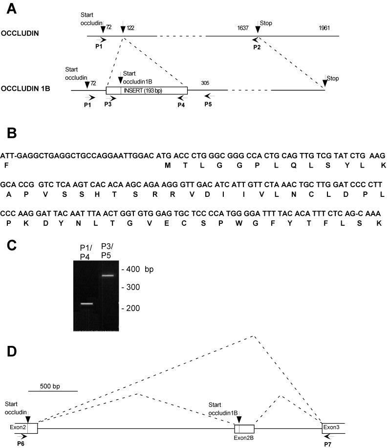
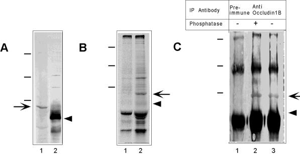
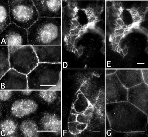
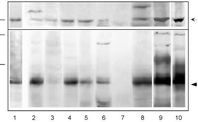
Occludin and claudin are the major integral membrane components of the mammalian tight junction. Although more than 11 distinct claudins have been identified, only 1 occludin transcript has been reported thus far. Therefore, we searched by reverse transcription-PCR for occludin-related sequences in Madin-Darby canine kidney (MDCK) mRNA and identified a transcript encoding an alternatively spliced form of occludin, designated occludin 1B. The occludin 1B transcript contained a 193-base pair insertion encoding a longer form of occludin with a unique N-terminal sequence of 56 amino acids. Analysis of the MDCK occludin gene revealed an exon containing the 193-base pair sequence between the exons encoding the original N terminus and the distal sequence, suggesting that occludin and occludin 1B arise from alternative splicing of one transcript. To assess the expression and distribution of occludin 1B, an antibody was raised against its unique N-terminal domain. Immunolabeling of occludin 1B in MDCK cells revealed a distribution indistinguishable from that of occludin. Furthermore, occludin 1B staining at cell-to-cell contacts was also found in cultured T84 human colon carcinoma cells and in frozen sections of mouse intestine. Immunoblots of various mouse tissues revealed broad coexpression of occludin 1B with occludin. The wide epithelial distribution and the conservation across species suggests a potentially important role for occludin 1B in the structure and function of the tight junction.


Figures

Figure 1
Figure 1
(A) Scheme of occludin and occludin 1B cDNA. Occludin 1B cDNA is aligned relative to the cDNA of occludin, and the positions of the primers used for PCR amplification (P1–P5) are indicated. The 193-bp exon specific for occludin 1B is inserted in the cDNA of occludin and is shown as a box. This exon contains the start for occludin 1B, as indicated, and encodes 56 amino acids. P1 and P2 indicate the positions of the primers used to clone the transcript for occludin 1B. (B) Nucleotide sequence of the exon specific for occludin 1B and the deduced amino acid sequence. (C) RT-PCR verification of expression of occludin 1B in MDCK cells as a transcript containing an insert contiguous at both ends with occludin mRNA. The products obtained with the primer pairs P1/P4 and P3/P5 (shown in Figure 1A) have the expected size and confirm the contiguity between occludin mRNA and the sequence shown in Figure 1B. Similar data were obtained with three separatemRNA preparations from MDCK cells. (D) Scheme of the structure of the canine occludin gene between exons 2 and 3. P6 and P7 indicate the positions of the primers used for the mapping of this portion of the gene. P6 is at the start of occludin (analogous to exon 2 in the mouse occludin gene), and P7 is on the previously designated exon 3 in the mouse gene. Downstream from exon 3, occludin and occludin 1B cDNAs are identical. The alternative start used for occludin 1B is contained in a novel exon (exon 2B) ∼700 bp upstream from exon 3.
Figure 2
Figure 2
Biochemical characterization of occludin 1B. (A) Immunoblot of extracts of MDCK cells with polyclonal anti-occludin 1B (lane 1) and anti-occludin (lane 2). No band was detected by the preimmune serum (not shown). The arrow and arrowhead indicate occludin 1B (∼70 kDa) and occludin (∼65 kDa), respectively. (B) SDS-PAGE and autoradiography of immunoprecipitates of extracts of MDCK cells. Cells metabolically labeled with [35S]methionine were immunoprecipitated with preimmune (lane 1) or anti-occludin 1B (lane 2) sera. The arrow points to the occludin 1B band. The arrowhead indicates the expected position of occludin. (C) Anti-occludin 1B immunoprecipitates were treated with (+) or without (−) alkaline phosphatase and then displayed by SDS-PAGE followed by Western blotting with anti-occludin. Anti-occludin 1B immunoprecipitated a specific band (∼70 kDa; arrow, lanes 2 and 3) not seen in the preimmune immunoprecipitate (lane 1) that was recognized by anti-occludin. Phosphatase treatment did not shift the position of this band. The expected position of occludin is indicated by the arrowhead. Molecular mass standards (dashes, from top to bottom) are 209, 125, and 78 kDa.
Figure 3
Figure 3
Indirect immunofluorescence with anti-occludin 1B (A, D, and F), preimmune occludin 1B (C), anti-occludin (B and E), and anti-myc (G). Specimens shown are MDCK cells (A–C and G), T84 cells (F), and mouse duodenum (D and E). (A and B) Double labeling of occludin 1B (A) and occludin (B) in MDCK cells. Note colocalization of the immune staining of both occludins at the cell-to-cell contacts. The nuclear labeling observed in A is nonspecific, because it is also detected by the preimmune serum (C). (D and E) Tangential section through the apical area of mouse intestinal epithelial cells double labeled with antiserum to occludin 1B (D) and with affinity-purified polyclonal anti-occludin (E). The staining for occludin 1B and occludin colocalized at the junctional complexes between intestinal epithelial cells. (F) T84 cells labeled with anti-occludin 1B. The staining is at the cell-to-cell contacts, most likely at the tight junction. (G) MDCK cells expressing myc-tagged full-length occludin 1B are labeled with anti-myc antibody. Transfected occludin 1B is targeted to the cell-to-cell contacts. Bars, 10 μm for A–C, F, and G; 5 μm for D and E.
Figure 4
Figure 4
Tissue expression of occludin 1B and occludin. Mouse organ homogenates were subjected to SDS-PAGE and immunoblotting with the use of anti-occludin 1B (top) or anti-occludin (bottom). The same analysis was done for extracts of MDCK cells and T84 human colon carcinoma cells. Protein loading for extracts from tissues and cell lines was 30 μg/lane, except for duodenum, for which the loading was 75 μg/lane. Molecular mass standards (dashes at left) are 78 kDa for the top panel and 125 and 78 kDa for the bottom panel. Occludin 1B and occludin are indicated by the arrow and the arrowhead, respectively. The lanes contain extracts from the following sources: mouse duodenum (1), brain (2), heart (3), kidney (4), liver (5), pancreas (6), spleen (7), testis (8), MDCK cells (9), and T84 cells (10).

References

    1. Ando-Akatsuka Y, Saitou M, Hirase T, Kishi M, Sakakibara A, Furuse M, Tsukita S. Interspecies diversity of the occludin sequence: cDNA cloning of human, mouse, dog, and rat-kangaroo homologues. J Cell Biol. 1996;133:43–47. - PMC - PubMed
    1. Balda MS, Anderson JM, Matter K. The SH3 domain of the tight junction protein ZO-1 binds to a serine protein kinase that phosphorylates a region C-terminal to this domain. FEBS Lett. 1996a;399:326–332. - PubMed
    1. Balda MS, Whitney JA, Flores C, Gonzalez S, Cereijido M, Matter K. Functional dissociation of paracellular permeability and transepithelial electrical resistance and disruption of the apical-basolateral intramembrane diffusion barrier by expression of a mutant tight junction membrane protein. J Cell Biol. 1996b;134:1031–1049. - PMC - PubMed
    1. Chen Y-H, Merzdorf C, Paul DL, Goodenough DA. COOH terminus of occludin is required for tight junction barrier function in early Xenopus embryos. J Cell Biol. 1997;138:891–899. - PMC - PubMed
    1. Cordenonsi M, Mazzon E, De Rigo L, Baraldo S, Meggio F, Citi S. Occludin dephosphorylation in early development of Xenopus laevis. J Cell Sci. 1997;110:3131–3139. - PubMed

Publication types

MeSH terms

LinkOut - more resources