. 2000 Feb;11(2):663-76.
doi: 10.1091/mbc.11.2.663.

Essential functions of protein tyrosine phosphatases PTP2 and PTP3 and RIM11 tyrosine phosphorylation in Saccharomyces cerevisiae meiosis and sporulation

Affiliations
Free PMC article

Essential functions of protein tyrosine phosphatases PTP2 and PTP3 and RIM11 tyrosine phosphorylation in Saccharomyces cerevisiae meiosis and sporulation

X L Zhan et al. Mol Biol Cell. 2000 Feb.
Free PMC article

Abstract

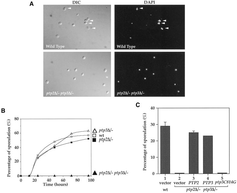
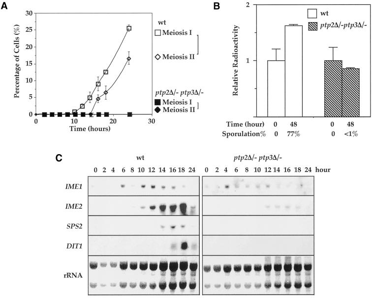
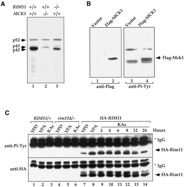
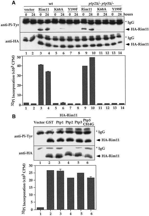
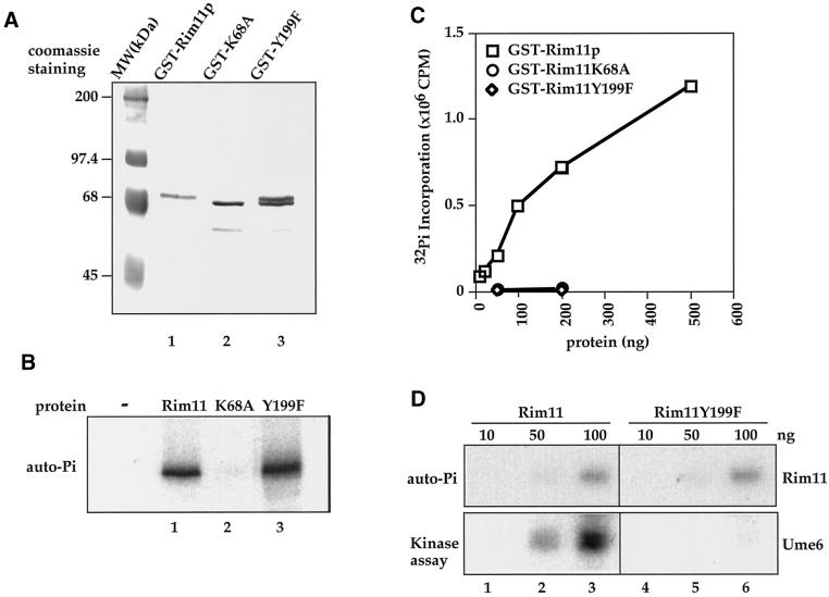
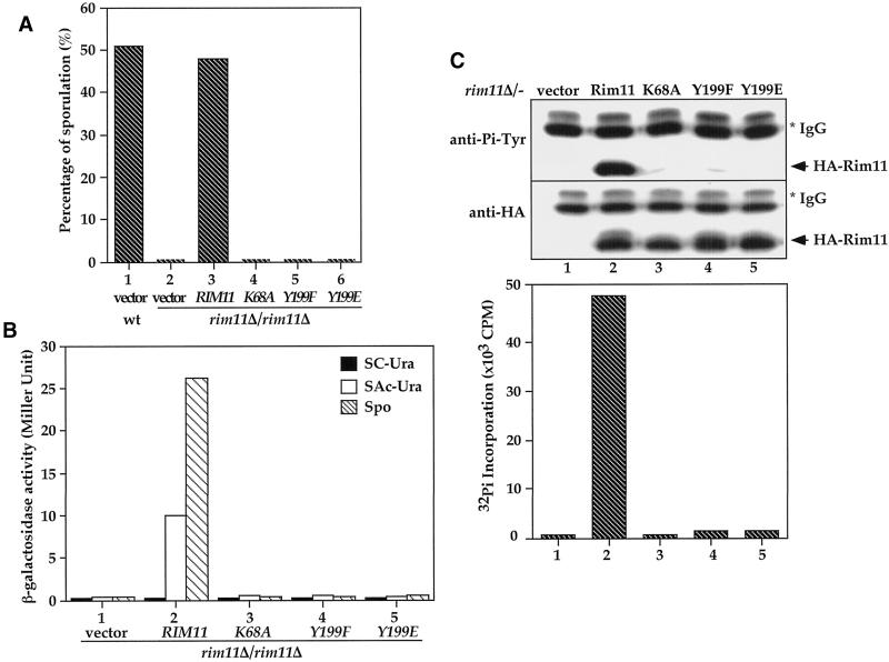
Tyrosine phosphorylation plays a central role in eukaryotic signal transduction. In yeast, MAP kinase pathways are regulated by tyrosine phosphorylation, and it has been speculated that other biochemical processes may also be regulated by tyrosine phosphorylation. Previous genetic and biochemical studies demonstrate that protein tyrosine phosphatases (PTPases) negatively regulate yeast MAP kinases. Here we report that deletion of PTP2 and PTP3 results in a sporulation defect, suggesting that tyrosine phosphorylation is involved in regulation of meiosis and sporulation. Deletion of PTP2 and PTP3 blocks cells at an early stage of sporulation before premeiotic DNA synthesis and induction of meiotic-specific genes. We observed that tyrosine phosphorylation of several proteins, including 52-, 43-, and 42-kDa proteins, was changed in ptp2Deltaptp3Delta homozygous deletion cells under sporulation conditions. The 42-kDa tyrosine-phosphorylated protein was identified as Mck1, which is a member of the GSK3 family of protein kinases and previously known to be phosphorylated on tyrosine. Mutation of MCK1 decreases sporulation efficiency, whereas mutation of RIM11, another GSK3 member, specifically abolishes sporulation; therefore, we investigated regulation of Rim11 by Tyr phosphorylation during sporulation. We demonstrated that Rim11 is phosphorylated on Tyr-199, and the Tyr phosphorylation is essential for its in vivo function, although Rim11 appears not to be directly regulated by Ptp2 and Ptp3. Biochemical characterizations indicate that tyrosine phosphorylation of Rim11 is essential for the activity of Rim11 to phosphorylate substrates. Our data demonstrate important roles of protein tyrosine phosphorylation in meiosis and sporulation


Figures

Figure 1
Figure 1
Diploid ptp2Δ/−ptp3Δ− double disruption cells are defective in sporulation. (A) PTP2 and PTP3 genes are required for sporulation. Wild-type (Y264, top) or ptp2Δ/ptp3Δ/− double deletion diploid (Y164, bottom) cells were sporulated in liquid sporulation media for 2 d. Cells were fixed, stained with DAPI, and examined by Normaski phase-contrast microscopy for spores (left panels) or fluorescence microscopy for DAPI-stained nuclei (right panels). A high proportion of wild-type cells have formed asci with a triad or tetrad of spores visible (top left panel, arrowheads) and these asci containing bi-, tri-, and tetranuclei (top right panel, arrowheads). No mature spores were found in ptp2Δ/ptp3Δ/− double deletion cells, and most of the cells only contain a single nucleus (lower right panel). (B) Defective sporulation in ptp2Δ/ptp3Δ/− double deletion cells but not single deletion of either phosphatase gene. Wild-type (Y264, open square), ptp2Δ/− (Y162, closed square), ptp3Δ/− (Y163, open triangle), or ptp2Δ/ptp3Δ/− (Y164, closed triangle) cells from liquid sporulation at the indicated time points were examined by phase-contrast microscopy. The percentage of sporulation was determined by counting the numbers of asci containing four mature spores. (C) Ptp3 Tyr-phosphatase activity is required for its function in sporulation. Wild-type (Y264, column 1) or ptp2Δ/ptp3Δ/− cells (Y164, column 2) harboring vector, single-copy PTP2 (pXZ223, column 3), PTP3 (pXZ209, column 4), or ptp3C814G (pXZ210, column 5), which encodes a catalytic-deficient phosphatase, were shifted to sporulation medium, and the sporulation efficiency was determined by counting the number of sporulated cells.
Figure 2
Figure 2
ptp2Δ/− ptp3Δ/- deletion cells were blocked before pre-meiotic DNA synthesis and induction of sporulation specific genes was decreased in double deletion cells. (A) ptp2Δ/ptp3Δ/− double deletion cells are arrested before meiosis I in sporulation medium. Wild-type (Y264) and ptp2Δ/ptp3Δ/− (Y164) cells were withdrawn from sporulation medium at the indicated time points. Cells were stained with DAPI and examined by fluorescence microscopy to determine the percentage of cells that had completed meiosis I or meiosis II. Tetranucleate cells (also include trinucleate cells) were counted as cells that completed meiosis II, whereas cells with more than one nucleus were counted as cells that completed meiosis I. Open square, meiosis I in wild type; open diamond, meiosis II in wild type; closed square, meiosis I in ptp2Δ/ptp3Δ/−; closed diamond, meiosis II in ptp2Δ/ptp3Δ/−, respectively. (B). Premeiotic DNA synthesis is absent in ptp2Δ/ptp3Δ/− double deletion cells. Wild-type diploid yeast cells (Y264, open bars) or ptp2Δ/ptp3Δ/− (Y164, closed bars) were grown in presporulation medium with [14C]uracil to label the endogenous pool of nucleotides. Cells were shifted into sporulation medium for 48 h in the absence of [14C]uracil. DNA was isolated, and total radioactivity incorporated in DNA was determined by scintillation counting. In parallel, the sporulation efficiency of the same cell samples was determined by microscopic examination. Average and SD from duplicated assays are shown. (C) Expression of sporulation-specific genes is decreased in ptp2Δ/ptp3Δ/− double deletion cells. RNA was prepared from either wild-type (Y264, left panels) or ptp2Δ/ptp3Δ/− double deletion cells (Y164, right panels) taken at various times after transferred to sporulation medium. Northern hybridization was performed on total RNA with probes derived from IME1, IME2, SPS2, and DIT1 genes as indicated. The amount of total RNA loaded in each lane was visualized with methylene blue staining for rRNA (bottom panels).
Figure 3
Figure 3
Elevated protein tyrosine phosphorylation in ptp2Δ/ptp3Δ/− double deletion cells during sporulation. (A) Tyrosine phosphorylation of 52-, 43-, and 42-kDa proteins is up-regulated in ptp2Δ/ptp3Δ/− double deletion cells during sporulation. Total cellular lysates were prepared from wild type (Y264, lanes 9–12), ptp2Δ/− single deletion (Y162, lanes 1–4), ptp3Δ/− single deletion (Y163, lanes 5–8), and ptp2Δ/ptp3Δ/− double deletion (Y164, lanes 13–16) grown in sporulation medium for various times. Equal amount of lysates were resolved on SDS-PAGE and subjected to immunoblotting with anti-Pi-Tyr antibody. The prominent tyrosine-phosphorylated proteins were denoted p42, p43, and p52 according to their apparent molecular masses. (B) Competition of anti-Pi-Tyr Western blots. Cell lysates prepared from wild-type (Y264) or ptp2Δ/ptp3Δ/− double deletion cells (Y164) incubated in sporulation medium for 24 h were subjected to anti-Pi-Tyr Western blotting (lanes 1 and 2). The immunoreactive signals were specifically competed if the anti-Pi-Tyr antibody was preincubated with 1 mM phosphotyrosine (lanes 3 and 4). As a control, no signal was detected on Western blotting without primary anti-Pi-Tyr antibody (lanes 5 and 6).
Figure 4
Figure 4
Tyrosine phosphorylation of Mck1 and Rim11. (A) Deletion of MCK1 and RIM11 in ptp2Δ/ptp3Δ/− double deletion cells eliminated Tyr phosphorylation of p42 and p43, respectively. Lysates were prepared from ptp2Δ/ptp3Δ/− (Y164, lane 1), ptp2Δ/ptp3Δ/mck1Δ/− (Y165, lane 2), and ptp2Δ/ptp3Δ/rim11Δ/− cells (Y166, lane 3) taken 24 h after being shifted to sporulation medium. Tyr phosphorylation was detected by immunoblotting with anti-Pi-Tyr antibody. (B) Flag-tagged MCK1 is phosphorylated on Tyr in vivo. Control vector (pRS424, lanes 1 and 3) or Flag epitope-tagged Mck1 (pRS424-Flag-MCK1, lanes 2 and 4) were introduced into ptp2Δ/ptp3Δ/− double deletion cells (Y164). Total cellular lysates were subjected to immunoblotting with anti-Flag (left, lanes 1 and 2) or anti-Pi-Tyr (right, lanes 3 and 4) antibody. Yeast cells containing epitope-tagged Mck1 showed an extra Tyr-phosphorylated band as indicated (Flag-Mck1). (C) Rim11 is phosphorylated on Tyr. Wild-type (lanes 1–3), rim11Δ/rim11Δ (KB268, lanes 4–6), or wild-type cells bearing an integrated copy of HA-RIM11 (KB600, lanes 7–14) were grown in YPD, YPA (rich acetate medium), or 1% KAc (sporulation medium). HA-Rim11 was immunoprecipitated from total cellular lysates and subjected to immunoblotting with anti-Pi-Tyr (top) or anti-HA (bottom) antibody to determine Tyr phosphorylation or protein levels, respectively.
Figure 5
Figure 5
Rim11 Tyr phosphorylation and activity are not affected by ptp2Δptp3Δ deletion or Ptp2 and Ptp3 overexpression. (A) Rim11 Tyr phosphorylation and kinase activity are not affected by ptp2Δ ptp3Δ deletion. Wild-type (Y264, lanes 1–8) or ptp2Δ/ptp3Δ/− double deletion cells (Y164, lanes 9–14) containing vector (pRS313, lanes 1–2), HA-tagged Rim11 wild type (pXZ352, lanes 3, 4, 9, and 10), K68A (pXZ353, lanes 5, 6, 11, and 12), or Y199F (pXZ354, lanes 7, 8, 13, and 14) were harvested at 0 and 24 h after being shifted to sporulation medium. HA-tagged proteins were immunoprecipitated and subjected to anti-Pi-Tyr (top) or anti-HA immunoblotting (middle) to determine Tyr phosphorylation or protein levels, respectively. In parallel, immunoprecipitated proteins were subjected to in vitro kinase assay using phospho-GS peptide as a substrate to determine the kinase activity (bottom). The radioactivity incorporated into the peptide was measured by scintillation counting. Average results from duplicated kinase assays are shown. (B) Overexpression of Ptp2 and Ptp3 has no effect on Rim11 Tyr phosphorylation and kinase activity. Wild-type cells (Y264) harboring HA-Rim11 (pXZ352) were transformed with vector (lane 1), pXZ134 (lane 2), pXZ110 (lane 3), pXZ113 (lane 4), pXZ123 (lane 5) or pXZ136 (lane 6) to overexpress GST or the indicated PTPases. Cellular lysates were prepared from transformants growing in SC-His,Ura, and HA-Rim11 was immunoprecipitated. Tyr phosphorylation (top), protein levels (middle), and kinase activities (bottom) were determined.
Figure 6
Figure 6
Tyrosine residue 199 in Rim11 is required for the recombinant kinase to phosphorylate substrates but not for autophosphorylation. (A) Coomassie blue staining of purified GST-Rim11, K68A and Y199F protein. GST fusion proteins were expressed in E. coli and purified by glutathione-agarose affinity chromatography and Mono-Q FPLC. (B) Autophosphorylation of GST-Rim11. Equal amounts of GST-Rim11 (lane 1), K68A (lane 2), and Y199F (lane 3) were incubated with [γ-32P]ATP to allow autophosphorylation. Proteins are analyzed on SDS-PAGE and followed by autoradiography. Rim11 and Y199F showed similar autophosphorylation activity, whereas no autophosphorylation was detected on K68A. (C) Kinase activity of recombinant GST-Rim11. The indicated amounts of purified GST fusion Rim11 wild type, K68A, and Y199F were used in in vitro kinase assays with phospho-GS peptide as an artificial substrate. The radioactivity incorporated into peptide was determined by scintillation counting. Square, activity of Rim11; circle, K68A; diamond, Y199F. (D) Tyr-199 is required for Rim11 to phosphorylate Ume6, a physiological substrate of Rim11. Recombinant GST-Ume6 was used as a substrate for GST-Rim11 (lanes 1–3) or GST-Rim11Y199F (lanes 4–6) in the in vitro kinase assay. GST-Ume6 was incubated with the indicated amount of GST-Rim11 or GST-Rim11Y199F in the presence of [γ-32P]ATP. The proteins were resolved by SDS-PAGE and subjected to autoradiography to visualize Rim11 autophosphorylation (top) or phosphorylation of Ume6 (bottom).
Figure 7
Figure 7
Tyrosine phosphorylation of Rim11 at the Tyr-199 residue is required for Rim11 kinase activity and its in vivo function. (A) Tyrosine phosphorylation and kinase activity are essential for RIM11 function in sporulation. Wild-type (KB600, column 1) cells harboring control vector or rim11Δ/rim11Δ deletion cells (KB268) containing vector (pRS426, column 2), HA-tagged wild-type RIM11 (pKB166, column 3), rim11K68A (pKB199, column 4), rim11Y199F mutant (pKB201, column 5), or rim11Y199E (pXZ356, column 6) were cultured in sporulation medium. The percentage of sporulated cells after 24 h was determined by microscopic counting. (B) Tyrosine phosphorylation and kinase activity are essential for Rim11 function in ime2-lacZ induction. An a/α rim11Δ/rim11Δ ime2-lacZ/IME2 diploid was transformed with vector (column 1), HA-tagged Rim11 wild-type (pKB166, column 2), K68A (pKB199, column 3), Y199F (pKB201, column 4), or Y199E mutant (pXZ356, column 5) and cultured in glucose and acetate or 8 h after cells were shifted to sporulation media. The expressions of ime2-lacZ in glucose (SC-Ura, closed bar), acetate (SAc-Ura, open bar), and sporulation medium (Spo, hatched bar) were determined by β-galactosidase assay. Activities are in Miller units and are averages of two duplicated assays. Y199F and Y199E mutations eliminate Rim11 in vivo kinase activity. HA-tagged Rim11 (pKB166), Rim11K68A (pKB199), Rim11Y199F (pKB201),or Rim11Y199E (pXZ356) was expressed in rim11Δ/rim11Δ deletion cells (KB268). The HA-tagged proteins were immunoprecipitated from yeast cell lysates with anti-HA antibody. The Tyr phosphorylation (top), protein levels (middle), and kinase activity (bottom) were determined by anti-Pi-Tyr, anti-HA immunoblotting, or in vitro kinase assay.

References

    1. Bowdish KS, Yuan HE, Mitchell AP. Analysis of RIM11, a yeast protein kinase that phosphorylates the meiotic activator IME1. Mol Cell Biol. 1994;14:7909–7919. - PMC - PubMed
    1. Cobb MH, Goldsmith EJ. How MAP kinases are regulated. J Biol Chem. 1995;270:14843–14846. - PubMed
    1. Dale TC. Signal transduction by the Wnt family of ligands. Biochem J. 1998;329:209–223. - PMC - PubMed
    1. Eisenmann DM, Kim SK. Signal transduction and cell fate specification during Caenorhabditis elegans vulval development. Curr Opin Genet Dev. 1994;4:508–516. - PubMed
    1. Friesen H, Lunz R, Doyle S, Segall J. Mutation of the SPS1-encoded protein kinase of Saccharomyces cerevisiae leads to defects in transcription and morphology during spore formation. Genes Dev. 1994;8:2162–2175. - PubMed

Publication types

MeSH terms