. 2000 Feb;11(2):773-93.
doi: 10.1091/mbc.11.2.773.

Identification of novel, evolutionarily conserved Cdc42p-interacting proteins and of redundant pathways linking Cdc24p and Cdc42p to actin polarization in yeast

Affiliations
Free PMC article

Identification of novel, evolutionarily conserved Cdc42p-interacting proteins and of redundant pathways linking Cdc24p and Cdc42p to actin polarization in yeast

E Bi et al. Mol Biol Cell. 2000 Feb.
Free PMC article

Abstract

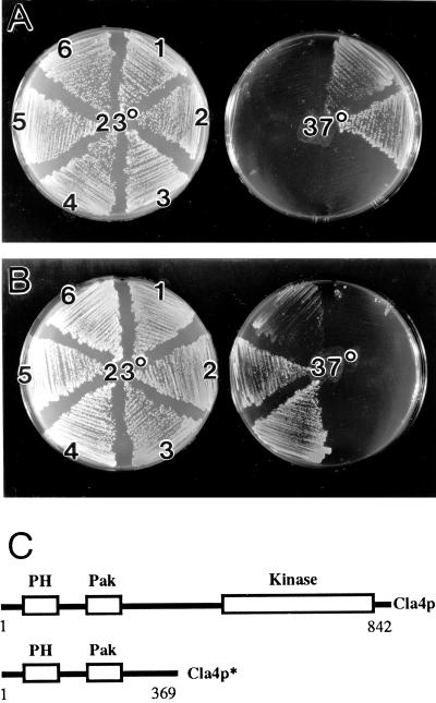
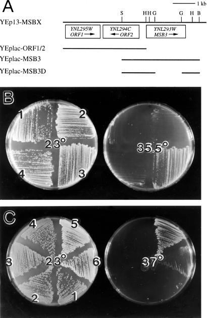
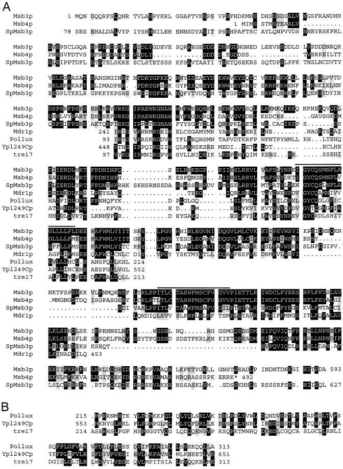
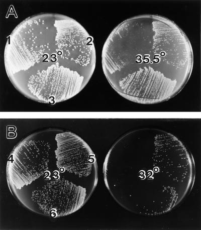
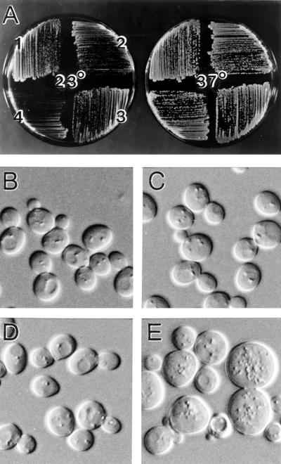
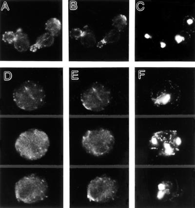
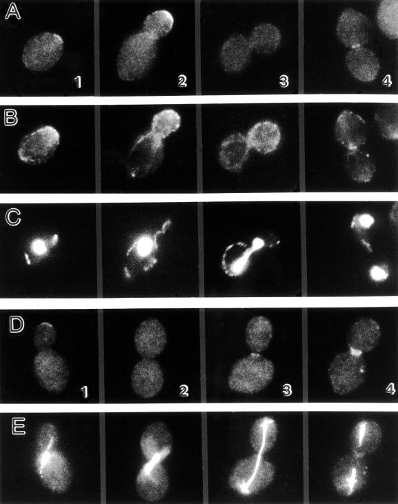
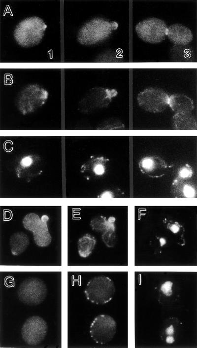
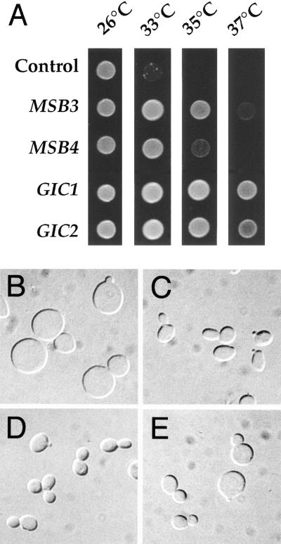
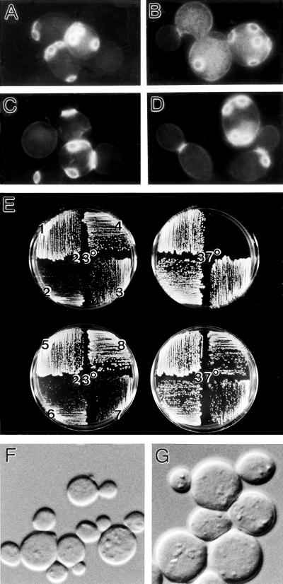
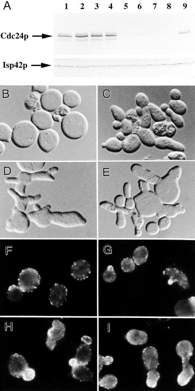
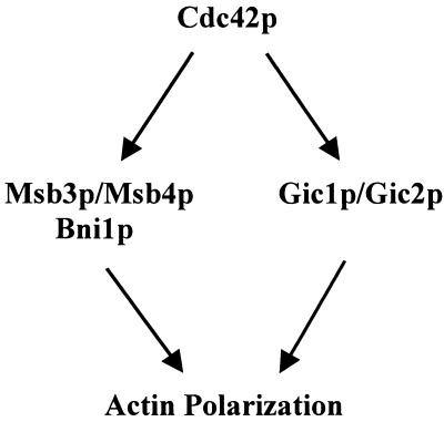
In the yeast Saccharomyces cerevisiae, Cdc24p functions at least in part as a guanine-nucleotide-exchange factor for the Rho-family GTPase Cdc42p. A genetic screen designed to identify possible additional targets of Cdc24p instead identified two previously known genes, MSB1 and CLA4, and one novel gene, designated MSB3, all of which appear to function in the Cdc24p-Cdc42p pathway. Nonetheless, genetic evidence suggests that Cdc24p may have a function that is distinct from its Cdc42p guanine-nucleotide-exchange factor activity; in particular, overexpression of CDC42 in combination with MSB1 or a truncated CLA4 in cells depleted for Cdc24p allowed polarization of the actin cytoskeleton and polarized cell growth, but not successful cell proliferation. MSB3 has a close homologue (designated MSB4) and two more distant homologues (MDR1 and YPL249C) in S. cerevisiae and also has homologues in Schizosaccharomyces pombe, Drosophila (pollux), and humans (the oncogene tre17). Deletion of either MSB3 or MSB4 alone did not produce any obvious phenotype, and the msb3 msb4 double mutant was viable. However, the double mutant grew slowly and had a partial disorganization of the actin cytoskeleton, but not of the septins, in a fraction of cells that were larger and rounder than normal. Like Cdc42p, both Msb3p and Msb4p localized to the presumptive bud site, the bud tip, and the mother-bud neck, and this localization was Cdc42p dependent. Taken together, the data suggest that Msb3p and Msb4p may function redundantly downstream of Cdc42p, specifically in a pathway leading to actin organization. From previous work, the BNI1, GIC1, and GIC2 gene products also appear to be involved in linking Cdc42p to the actin cytoskeleton. Synthetic lethality and multicopy suppression analyses among these genes, MSB, and MSB4, suggest that the linkage is accomplished by two parallel pathways, one involving Msb3p, Msb4p, and Bni1p, and the other involving Gic1p and Gic2p. The former pathway appears to be more important in diploids and at low temperatures, whereas the latter pathway appears to be more important in haploids and at high temperatures.


Figures

Figure 1
Figure 1
Suppression of cdc24ts and cdc42ts mutations by multicopy MSB1 or multicopy truncated CLA4. The transformants described below were streaked onto SC-Leu-Ura plates with 1 M sorbitol (A) or SC-Leu plates with 1 M sorbitol (B) and incubated at the indicated temperatures. (A) cdc24-11 strain YEF319 was transformed with (1) YEp352-CDC42 and YEp13-CLA4*, (2) YEp352-CDC42 and YEp13-MSB1, (3) YEp13-CLA4* and pPB191 (high-copy MSB1), (4) YEp352-CDC42 and YEp13, (5) YEp24 and YEp13-CLA4*, or (6) YEp24 and YEp13-MSB1. (B) cdc24-12 strain YEF323 (1–3) and cdc42-1 strain DJTD2–16D (4–6) were transformed with (3 and 6) YEp13, (2 and 5) YEp13-CLA4*, or (1 and 4) YEp13-MSB1. (C) Structures of full-length Cla4p and of the truncated Cla4p encoded by plasmid YEp13-CLA4*.
Figure 2
Figure 2
Suppression of cdc24ts and cdc42ts mutations by multicopy MSB3. (A) Physical maps of the inserts in plasmid YEp13-MSBX and in the subclones tested for suppression. All HindIII (H), SacI (S), BglII (G), and BamHI (B) sites are shown. (B and C) The transformants described below were streaked onto SC-Leu-Ura plates with 1 M sorbitol (B) or SC-Leu plates with 1 M sorbitol (C) and incubated at the indicated temperatures. (B) cdc24-11 strain YEF319 harboring plasmid YEp352-CDC42 was transformed with (1) YEp13, (2) YEplac-MSB3D, (3) YEplac-MSB3, or (4) YEplac-ORF1/2. (C) cdc24-13 strain YEF327 (1 and 2), cdc24-12 strain YEF323 (3 and 4), and cdc42-1 strain DJTD2–16D (5 and 6) were transformed with (2, 4, and 6) YEp13 or (1, 3, and 5) YEp13-MSBX.
Figure 3
Figure 3
Alignment of Msb3p-homologous sequences from different organisms using the Genetics Computer Group (Madison, WI) Pile-Up program plus some manual adjustments. (A) The complete sequences of Msb3p, Msb4p, and SpMsb3p are shown except for the COOH terminus of Msb3p and the NH2 and COOH termini of SpMsb3p; for the other proteins, only the regions of clear homology to Msb3p are shown (213 amino acids for Mdr1p; 105–122 amino acids for Pollux, Ypl249Cp, and tre17). Black boxes indicate amino acids identical to those in Msb3p; gray boxes indicate amino acids similar to those in Msb3p (D/E; Q/N; R/K; S/T; I/L/V). Overlining indicates the possible transmembrane domains (see text). The proteins depicted (with accession numbers) include the four proteins from S. cerevisiae (Msb3p [U23084], Msb4p [S51885], Mdr1p [Z72885], and Ypl249Cp [Z67751]); one from S. pombe (SpMsb3p [the product of a previously uncharacterized ORF on cosmid SPCC4G3 from chromosome III]); one from D. melanogaster (Pollux [U50542]); and one from H. sapiens (tre17 [S22155]). Other known proteins showing this region of homology include one from S. pombe (Z56276); seven from C. elegans (Z78539, U67954, Q09445, U49940, U42831, U41540, and U29244); one from M. musculus (Tbc1 [U33005]); and one from H. sapiens (D13644). (B) The sequences of Pollux, Ypl249Cp, and tre17 are shown for the region immediately downstream of the portions of these proteins shown in A. Black boxes indicate amino acids identical in two or more of the proteins; gray boxes indicate amino acids similar (as in A) in two or more of the proteins.
Figure 4
Figure 4
Suppression of cdc24-11 by multicopy MSB4. Strain YEF319 with (A) or without (B) plasmid YEp352-CDC42 was transformed with plasmid YEp13 (1 and 4), YEplac-MSB4 (2 and 5), or YEplac-MSB3 (3 and 6). The transformants were streaked onto SC-Leu-Ura plates with 1 M sorbitol (A) or SC-Leu plates with 1 M sorbitol (B) and incubated at the indicated temperatures.
Figure 5
Figure 5
Phenotypic consequences of deleting MSB3 and MSB4. (A) Growth of single and double mutants on YPD solid medium at 23 or 37°C. (B–E) Morphology of the single and double mutants in YPD liquid medium at 23°C, as observed by DIC microscopy. (A, sector 1, and B) YEF473A (wild-type); (A, sector 2, and C) YEF1239 (msb3Δ::HIS3); (A, sector 3, and D) YEF1247 (msb4Δ::HIS3); (A, sector 4, and E) YEF1269 (msb3Δ::HIS3 msb4Δ::HIS3). B–E are printed at the same magnification.
Figure 6
Figure 6
Actin and septin organization in the msb3 msb4 double mutant. Cells of wild-type strain YEF473A (A–C) and msb3 msb4 double-mutant strain YEF1269 (D–F) growing exponentially in YPD liquid medium at 23°C were triple stained for actin (A and D), Cdc11p (B and E), and DNA (C and F). All panels are printed at the same magnification.
Figure 7
Figure 7
Immunolocalization of HA-tagged Msb3p. Cells of strain YEF1474 harboring plasmid YEplac-3HA-MSB3 growing exponentially in SC-Leu liquid medium at 23°C were triple stained for HA-tagged Msb3p (A), actin (B), and DNA (C) or double stained for HA-tagged Msb3p (D) and tubulin (E). Individual cells are numbered for reference in the text.
Figure 8
Figure 8
Immunolocalization of HA-tagged Msb4p (A–C) and dependence of Msb3p localization on Cdc42p (D–I). (A–C) Cells of strain YEF1475 containing plasmid YEplac-3HA-MSB4 growing exponentially in SC-Leu liquid medium at 23°C were triple stained for HA-tagged Msb4p (A), actin (B), and DNA (C). Individual cells are numbered for reference in the text. (D–I) Cells of strain YEF1517 (temperature-sensitive for Cdc42p function; see text) harboring plasmid YEplac-3HA-MSB3 growing exponentially in SC-Leu liquid medium at 23°C (D–F) or shifted to 37°C for 4 h (G–I) were triple stained for HA-tagged Msb3p (D and G), actin (E and H), and DNA (F and I).
Figure 9
Figure 9
Suppression of the gic1 gic2 double mutant by multicopy MSB3 or MSB4. (A) Suspensions of strain CCY1024–19C harboring plasmid pSM217 (control), pCC1107 (MSB3), pCC1108 (MSB4), pCC904 (GIC1), or pCC967 (GIC2) were spotted on YPD plates and incubated for 2 days at the indicated temperatures. (B–E) Cells of strain CCY1024–19C containing plasmid pSM217 (B), pCC904 (C), pCC1107 (D), or pCC1108 (E) were grown to exponential phase in SD+Ade+Trp+casamino acids medium at 26°C and then shifted to 37°C for 4 h before fixation and examination by DIC microscopy. B–E are printed at the same magnification.
Figure 10
Figure 10
Comparison of msb3 msb4, bni1, and gic1 gic2 mutant phenotypes. (A–D) Cells of diploid wild-type strain YEF473 (A), msb3Δ::HIS3/msb3Δ::HIS3 msb4Δ::TRP1/msb4Δ::TRP1 strain YEF1631 (B), bni1Δ::HIS3/bni1Δ::HIS3 strain HH799 (C), and gic1-Δ1::LEU2/ gic1-Δ1::LEU2 gic2-Δ2::TRP1/gic2-Δ2::TRP1 strain YEF1662 (D) were grown to exponential phase in YM-P liquid medium at 23°C, then fixed and stained with Calcofluor. (E) Haploid wild-type strain YEF473A (1), msb3Δ::HIS3 msb4Δ::HIS3 strain YEF1269 (2), bni1Δ::HIS3 strain YJZ426 (3), gic1-Δ1::LEU2 gic2-Δ2::TRP1 strain CCY1042–12B (4), and diploid strains YEF473 (5), YEF1631 (6), HH799 (7), and YEF1662 (8) were streaked onto YPD plates and incubated for 2 days at the indicated temperatures. (F and G) Cells of msb3 msb4 double-mutant haploid strain YEF1269 (F) and of double-mutant diploid strain YEF1631 (G) were grown to exponential phase in YM-P liquid medium at 23°C and examined by DIC microscopy. A–D, F, and G are printed at the same magnification.
Figure 11
Figure 11
Polarity establishment in the absence of Cdc24p. (A) Western-blot analysis using antibodies to Cdc24p and (as a control) the mitochondrial outer membrane protein Isp42p. (B–E) Cell morphologies as observed by DIC microscopy. (F–I) Immunolocalization of actin using anti-actin antibodies. Strain YEF1201 (cdc24Δ::HIS3 [pMGF5]) was transformed with plasmids YEp352-CDC42 and YEp13 (A, lanes 1 and 5, B, and F), YEp352-CDC42 and YEp13-CLA4* (A, lanes 2 and 6, C, and G), YEp352-CDC42 and YEp13-MSB1 (A, lanes 3 and 7, D, and H), or YEp352–42CLA4* and YEp13-MSB1 (A, lanes 4 and 8, E, I). Cells were grown to exponential phase at 30°C under mildly inducing conditions for GAL1-CDC24 (SC-Leu-Ura liquid medium containing 2% glucose plus 2% galactose), and samples were removed for immunoblot analysis (A, lanes 1–4). Cells were then shifted to repressing conditions (SC-Leu-Ura liquid medium containing 2% glucose only) for 12 h, diluted further with the same medium, and incubated for an additional 4 h before harvesting for immunoblot (A, lanes 5–8) and microscopic (B–I) analysis. Wild-type strain YEF473 growing exponentially in liquid SC medium at 30°C was used as a control (A, lane 9). B–I are printed at the same magnification.
Figure 12
Figure 12
Parallel pathways for signaling from Cdc42p to the actin cytoskeleton. See text for details.

References

    1. Adams AEM, Johnson DI, Longnecker RM, Sloat BF, Pringle JR. CDC42 and CDC43, two additional genes involved in budding and the establishment of cell polarity in the yeast Saccharomyces cerevisiae. J Cell Biol. 1990;111:131–142. - PMC - PubMed
    1. Adams AEM, Pringle JR. Relationship of actin and tubulin distribution to bud growth in wild-type and morphogenetic-mutant Saccharomyces cerevisiae. J Cell Biol. 1984;98:934–945. - PMC - PubMed
    1. Adams AEM, Pringle JR. Staining of actin with fluorochrome-conjugated phalloidin. Methods Enzymol. 1991;194:729–731. - PubMed
    1. Aghazadeh B, Zhu K, Kubiseski TJ, Liu GA, Pawson T, Zheng Y, Rosen MK. Structure and mutagenesis of the Dbl homology domain. Nat Struct Biol. 1998;5:1098–1107. - PubMed
    1. Amatruda JF, Cooper JA. Purification, characterization, and immunofluorescence localization of Saccharomyces cerevisiae capping protein. J Cell Biol. 1992;117:1067–1076. - PMC - PubMed

Publication types

MeSH terms

Substances